. 2010 Feb;24(2):415-24.
doi: 10.1096/fj.09-140566. Epub 2009 Oct 2.

Ca2+-independent positive molecular inotropy for failing rabbit and human cardiac muscle by alpha-myosin motor gene transfer

Affiliations

Ca2+-independent positive molecular inotropy for failing rabbit and human cardiac muscle by alpha-myosin motor gene transfer

Todd J Herron et al. FASEB J. 2010 Feb.

Abstract

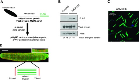
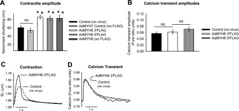
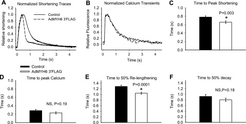
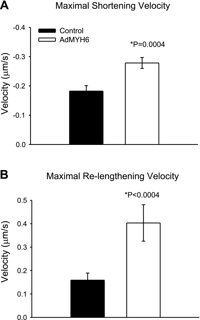
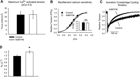
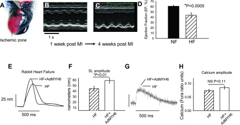
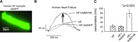
Current inotropic therapies used to increase cardiac contractility of the failing heart center on increasing the amount of calcium available for contraction, but their long-term use is associated with increased mortality due to fatal arrhythmias. Thus, there is a need to develop and explore novel inotropic therapies that can act via calcium-independent mechanisms. The purpose of this study was to determine whether fast alpha-myosin molecular motor gene transfer can confer calcium-independent positive inotropy in slow beta-myosin-dominant rabbit and human failing ventricular myocytes. To this end, we generated a recombinant adenovirus (AdMYH6) to deliver the full-length human alpha-myosin gene to adult rabbit and human cardiac myocytes in vitro. Fast alpha-myosin motor expression was determined by Western blotting and immunocytochemical analysis and confocal imaging. In experiments using electrically stimulated myocytes from ischemic failing hearts, AdMYH6 increased the contractile amplitude of failing human [23.9+/-7.8 nm (n=10) vs. AdMYH6 amplitude 78.4+/-16.5 nm (n=6)] and rabbit myocytes. The intracellular calcium transient amplitude was not altered. Control experiments included the use of a green fluorescent protein or a beta-myosin heavy chain adenovirus. Our data provide evidence for a novel form of calcium-independent positive inotropy in failing cardiac myocytes by fast alpha-myosin motor protein gene transfer.


Figures

Figure 1
Figure 1
AdMYH6 gene transfer in healthy β-MyHC-dominant rabbit cardiac myocytes. A) A recombinant adenovirus (AdMYH6) was designed to deliver and express the full-length human α-MyHC fast myosin motor in adult cardiac myocytes. B) Western blotting shows that α-MyHC expression was detectable 24 h after viral gene transfer and increased 48 h after gene transfer. C) Immunocytochemical analysis and low magnification (×20) demonstrate highly efficient (∼98%) gene transfer using AdMYH6. Green fluorescence represents the presence of the FLAG epitope; nuclear staining is blue (DAPI). D) Immunocytochemical analysis and high-resolution confocal imaging demonstrate that α-MyHC protein was expressed homogenously across the length and width of cardiac myocytes (whole-cell image, scale bar=20 μm) and incorporated appropriately in the sarcomere A band (inset, scale bar=5 μm) in between the Z lines (red, α-actinin).
Figure 2
Figure 2
Functional effects of AdMYH6 gene transfer on healthy rabbit cardiac myocytes. AdMYH6 gene transfer potentiates sarcomere shortening, whereas the calcium transient is unaltered. All experiments were performed 48 h after gene transfer. A) Summary of AdMYH6 effects on contraction amplitude. AdMYH7 control values did not differ from nontransduced control values (53.9±5.23 nm, n=18 vs. 60.5±4.5 nm, n=74). AdMYH6 increased contraction amplitude regardless of the presence or location of the FLAG epitope. AdMYH6 3′FLAG amplitude = 85.8 ± 4.8 nm, n = 86; AdMYH6 5′FLAG amplitude = 83.1 ± 5.23 nm, n = 37; AdMYH6 no FLAG amplitude = 82.3 ± 7.7 nm, n = 19. B) Calcium transient amplitudes were the same between nontransduced control (0.057±0.005, n=38), AdMYH6 3′FLAG-treated (0.062±0.006, n=34), and AdMYH6 5′FLAG-treated (0.069±0.006, n=32) cardiac myocytes. C, D) Representative recordings of sarcomere length (SL) shortening (C) and intracellular calcium transients (D). *P < 0.05 vs. control (no virus); P < 0.05 vs. AdMYH7 control; ANOVA with Tukey post hoc between-group comparisons.
Figure 3
Figure 3
α-MyHC gene transfer speeds healthy rabbit myocyte shortening and relengthening kinetics independent of the calcium transient dynamics. A) Normalized sarcomere length-shortening traces show that AdMYH6 gene transfer sped sarcomere shortening and relengthening. B) Normalized intracellular calcium transient traces show that the kinetics of calcium release and decay were unaffected by AdMYH6 gene transfer. C) Time to peak of sarcomere shortening was faster in AdMYH6-transduced myocytes (0.66±0.03 s, n=86) than in nontransduced myocytes (0.78±0.03 s, n=74). D) Time to peak of the calcium transient, however, did not differ between the two groups (0.28±0.03 s, n=38; 0.23±0.03 s, n=34). E) Time to 50% relaxation was reduced by AdMYH6 gene transfer compared with that in nontransduced control myocytes. F) Time to 50% decay of the calcium transient was not affected. All values are means ± se. *P < 0.05 vs. control, two-tailed t test. Actual P values are shown in figure.
Figure 4
Figure 4
AdMYH6 gene transfer speeds the maximal rates of sarcomere shortening and relengthening in healthy β-MyHC-dominant rabbit myocytes. A) Comparison of maximal rate of sarcomere shortening in control and AdMYH6-treated myocytes. B) Comparison of maximal rate of sarcomere relengthening in control (n=74) and AdMYH6-treated myocytes (n=86). *P < 0.05; unpaired t test.
Figure 5
Figure 5
AdMYH6 effects on rabbit cardiac myocyte maximal calcium-activated (pCa 4.5) isometric contractile function. A) Maximal isometric tension generation of skinned (1% Triton X-100) cardiac myocytes was not affected by AdMYH6 gene transfer. B) Myofilament calcium sensitivity did not differ between control (•) and AdMYH6-treated (○) myocytes. P, tension development; P0, maximal tension development; P/P0, relative tension. C, D) Maximal rate of isometric cross-bridge cycling was faster in AdMYH6-treated cardiac myocytes vs. control myocytes (1.40±0.07 s−1, n=16 vs. 1.16±0.07 s−1, n=16). All values are means ± se. *P < 0.05 vs. control; two-tailed t test.
Figure 6
Figure 6
α-MyHC gene transfer increases the contractility of failing rabbit cardiac myocytes. A) An ischemic model of heart failure was implemented in adult rabbits. Successful coronary occlusion was confirmed by retrograde perfusion of Evans Blue dye. B, C) Representative m-mode echocardiography images of animals with a nonfailing heart (NF; B) and heart failure (HF; C) show that cardiac function was severely depressed after chronic LCX occlusion. D) Ventricular ejection fraction was depressed in HF vs. NF animals (43.9±2.2%, n=4 vs. 60.6±1.2%, n=4). E) Representative sarcomere length (SL) shortening measurements in HF cardiac myocytes 2 d after in vitro gene transfer with AdMYH6 (HF+AdMYH6) or with no virus treatment (HF). F) Summary data show that α-MyHC gene transfer increased SL shortening amplitude in electrically paced (1 Hz) single myocytes. HF amplitude = 44.6 ± 4.0 nm, n = 59; HF + AdMYH6 amplitude = 59.2 ± 4.5 nm, n = 50. G) Representative calcium transient recordings show no effect of AdMYH6 gene transfer on intracellular calcium cycling. H) Summary data show that the intracellular calcium transient amplitude was not significantly affected by α-MyHC gene transfer. HF amplitude = 0.074 ± 0.004, n = 53; HF + AdMYH6 amplitude = 0.085 ± 0.005 ratio units, n = 48.
Figure 7
Figure 7
AdMYH6 gene transfer augments failing human cardiac myocyte contraction. A) Fluorescent image of a single human cardiac myocyte demonstrates successful adenovirus-mediated gene transfer of GFP. Image was taken 48 h after gene transfer. B) Representative sarcomere length shortening measurements in human failing ventricular cardiac myocytes. α-MyHC gene transfer enhanced the contractile amplitude of failing human cardiac myocytes paced at 1 Hz. C) Summary of data. HF amplitude = 23.9 ± 7.8 nm, n = 10; HF+AdGFP amplitude = 21.8 ± 5.2nm, n = 5; HF + AdMYH6 amplitude = 78.4 ± 16.5 nm, n = 6. All values are means ± se. *P < 0.05 vs. control, two-tailed t test. Actual P values are shown in figure. Human myocyte data were analyzed by 1-way ANOVA as there were 3 groups to compare: HF, HF + AdGFP, and HF + AdMYH6.

References

    1. McKinsey T. A., Olson E. N. Toward transcriptional therapies for the failing heart: chemical screens to modulate genes. J Clin Invest. 2005;115:538–546. - PMC - PubMed
    1. Perrone S. V., Kaplinsky E. J. Calcium sensitizer agents: a new class of inotropic agents in the treatment of decompensated heart failure. Int J Cardiol. 2005;103:248–255. - PubMed
    1. Cohn J. N., Goldstein S. O., Greenberg B. H., Lorell B. H., Bourge R. C., Jaski B. E., Gottlieb S. O., McGrew F., 3rd, DeMets D. L., White B. G. A dose-dependent increase in mortality with vesnarinone among patients with severe heart failure: Vesnarinone Trial Investigators. N Engl J Med. 1998;339:1810–1816. - PubMed
    1. Antoniades C., Tousoulis D., Koumallos N., Marinou K., Stefanadis C. Levosimendan: beyond its simple inotropic effect in heart failure. Pharmacol Ther. 2007;114:184–197. - PubMed
    1. Cleland J. G. F., Freemantle N., Coletta A. P., Clark A. L. Clinical trials update from the American Heart Association: REPAIR-AMI, ASTAMI, JELIS, MEGA, REVIVE-II, SURVIVE, and PROACTIVE. Eur J Heart Fail. 2006;8:105–110. - PubMed