. 2009 Oct 15;183(8):4975-83.
doi: 10.4049/jimmunol.0901243.

The E3 ubiquitin ligase Cbl-b regulates expansion but not functional activity of self-reactive CD4 T cells

Affiliations

The E3 ubiquitin ligase Cbl-b regulates expansion but not functional activity of self-reactive CD4 T cells

Marie-Clare St Rose et al. J Immunol. .

Abstract

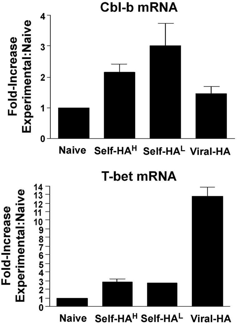
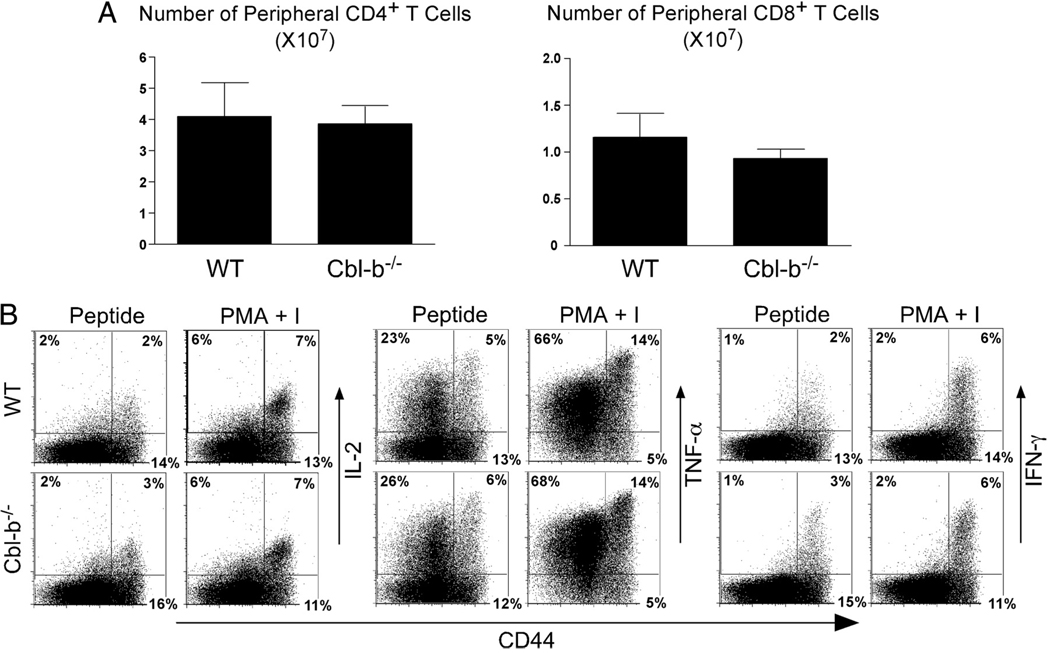
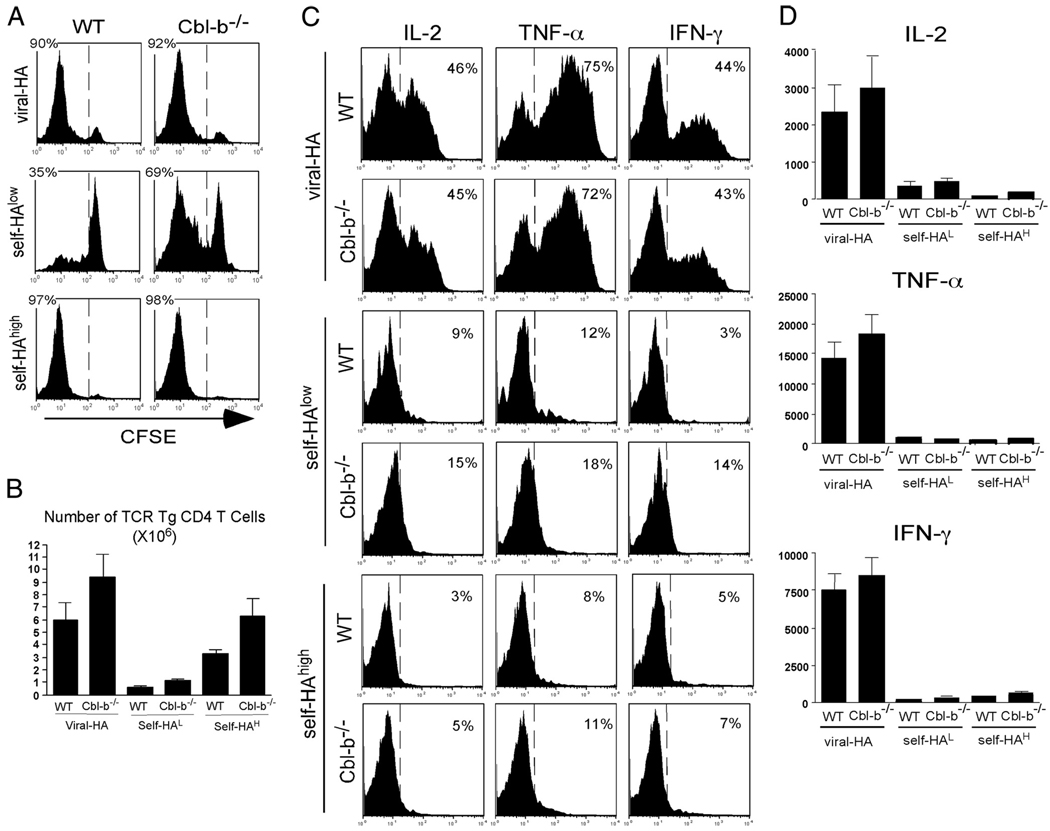
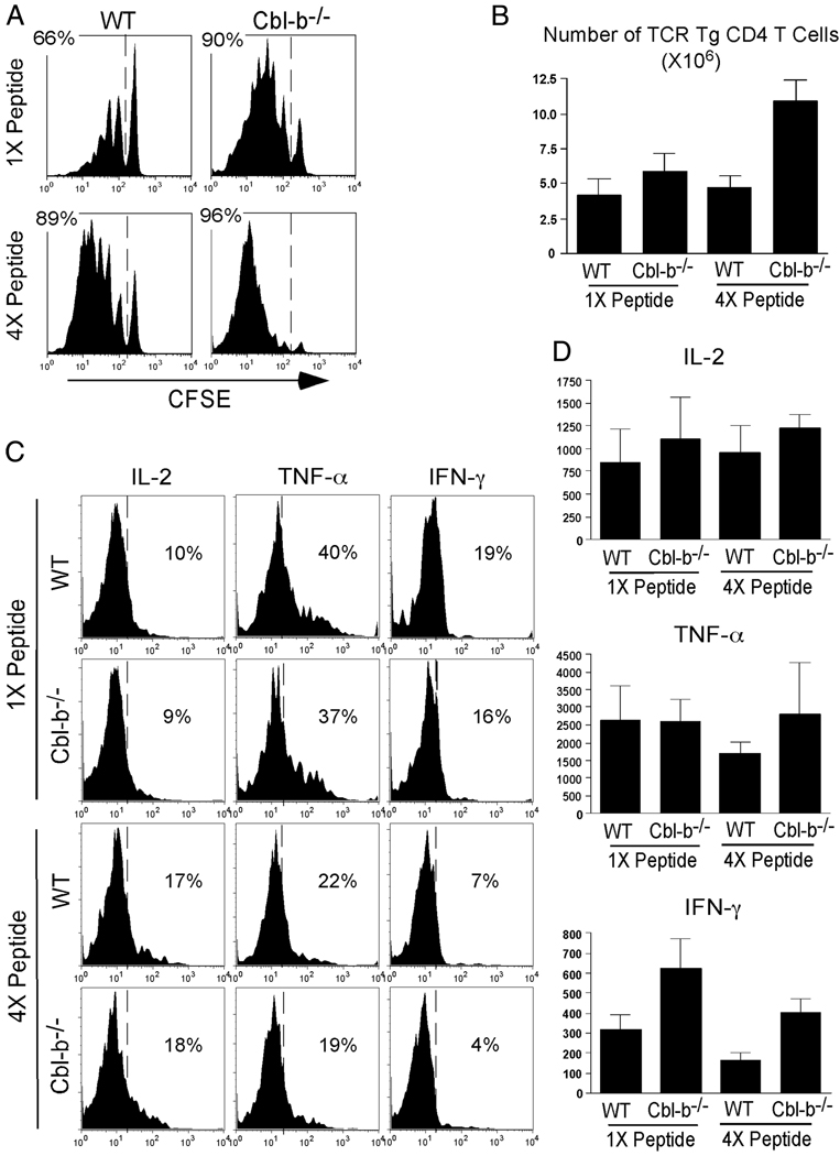
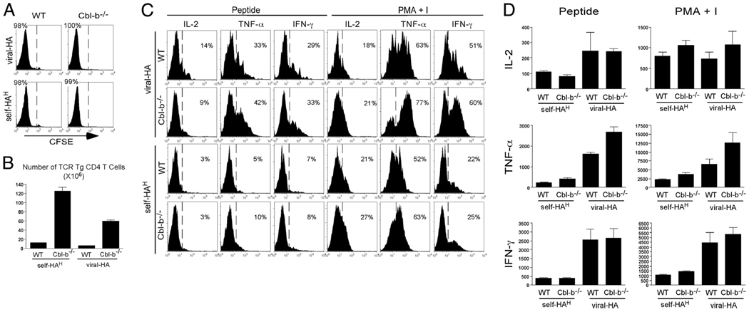
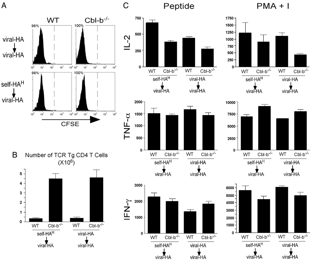
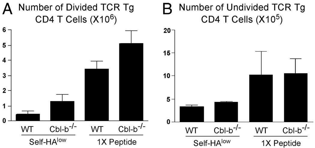
Cbl-b is an E3 ubiquitin ligase that limits Ag responsiveness in T cells by targeting TCR-inducible signaling molecules. Cbl-b deficiency thus renders T cells hyperresponsive to antigenic stimulation and predisposes individuals toward developing autoimmunity. In part because Cbl-b(-/-) T cells do not require CD28 costimulation to become activated, and insufficient costimulation is a critical parameter that confers anergy induction over effector differentiation, it has been hypothesized that Cbl-b(-/-) T cells are resistant to anergy. This possibility has been supported in models in which anergy is normally induced in vitro, or in vivo following exposure to soluble Ag boluses. In the current study, we characterized the response of Cbl-b(-/-) CD4 T cells in an in vivo system in which anergy is normally induced by a constitutively expressed peripheral self-Ag. Cbl-b expression increased in self-Ag-induced anergic wild-type CD4 T cells, and Cbl-b(-/-) CD4 T cells underwent more robust proliferation and expansion upon initially encountering cognate self-Ag compared with wild-type counterparts. Nevertheless, both wild-type and Cbl-b(-/-) CD4 T cells ultimately developed the same impaired ability to respond to antigenic restimulation. The more extensive expansion that occurred during the initial induction of anergy did, however, allow the anergic CD4 T cells to expand to greater numbers when they were functionally resuscitated following replacement of the initial source of tolerizing self-Ag with a viral form of the same Ag.


Conflict of interest statement

Disclosures

The authors have no financial conflict of interest.

Figures

FIGURE 1
FIGURE 1
Cbl-b mRNA expression increases in self-HA-tolerized TCR Tg CD4 T cells. Cbl-b and T-bet mRNAs were measured by quantitative real-time RT-PCR in TCR Tg CD4 T cell samples purified from spleens of viral-HA (n = 9), self-HAlow (self-HAL, n = 3, each determination deriving from 3 pooled recipient samples), and self-HAhigh (self-HAH, n = 12) recipients 6 days following adoptive transfer. (Note that these are the same samples that were analyzed for T-bet and IL-12Rβ2 mRNA expression in Fig. 5, C and D, of Ref. .) Data are expressed as the ratio relative to naive TCR Tg CD4 T cells.
FIGURE 2
FIGURE 2
TCR Tg Cbl-b−/− peripheral T cells are phenotypically and functionally similar to Cbl-b+/+ (WT) counterparts. A, The total number of CD4+ and CD8+ cells in pooled spleen and peripheral lymph nodes from 4-mo-old Cbl-b−/− and WT TCR Tg mice (corresponding to the donors used in Figs. 3–5). B, Cbl-b−/− and WT CD4+ T cells were stimulated with HA peptide or PMA plus I and then stained for surface CD44 and intracellular IL-2, TNF-α, or IFN-γ. Data shown are representative of three independent experiments.
FIGURE 3
FIGURE 3
Functional response of Cbl-b−/− CD4 T cells exposed to cognate self-Ag. CFSE-labeled Cbl-b−/− (Thy1.1/1.2) and WT (Thy1.1/1.1) TCR Tg CD4 T cells were adoptively cotransferred (5 × 10⁵ each) into viral-HA, self-HAlow, and self-HAhigh recipients and then recovered from spleens on day 4. A, Representative histograms of CFSE dilution. B, Expansion is plotted as the total number of TCR Tg CD4 T cells. C, Representative plots of intracellular IL-2, TNF-α, and IFN-γ expression following restimulation with HA peptide. D, Quantitation of cytokine expression corresponding to C. Total intracellular cytokine is expressed in arbitrary units, and was calculated by multiplying the percentage of cytokine expressing TCR Tg CD4 T cells by the corresponding mean fluorescence intensity values. n = 3 for each group, and all quantitative data are expressed as the mean ± SEM.
FIGURE 4
FIGURE 4
Cbl-b deficiency augments CD4 T cell expansion to cognate soluble peptide, but does not influence functional activity. CFSE-labeled Cbl-b−/− (Thy1.1/1.2) and WT (Thy1.1/1.1) TCR Tg CD4 T cells were adoptively cotransferred (5 × 10⁵ each) into NT recipients treated with boluses of soluble HA peptide given either once on day 0 (1×) or daily from days 0 to 3 (4×), and then recovered from spleens on day 4. A, Representative histograms of CFSE dilution. B, Expansion is plotted as the total number of TCR Tg CD4 T cells. C, Representative plots of intracellular IL-2, TNF-α, and IFN-γ expression following restimulation with HA peptide. D, Total cytokine expression corresponding to C. n = 3 for each group.
FIGURE 5
FIGURE 5
Cbl-b deficiency allows self-reactive CD4 T cells to massively expand, but does not confer resistance to anergy. CFSE-labeled Cbl-b−/− (Thy1.1/1.1) and WT (Thy1.1/1.2) TCR Tg CD4 T cells were adoptively cotransferred (5 × 10⁵ each) into viral-HA and self-HAhigh recipients and then recovered from spleens on day 4. A, Representative histograms of CFSE dilution. B, Expansion is plotted as the total number of TCR Tg CD4 T cells. C, Representative plots of intracellular IL-2, TNF-α, and IFN-γ expression following restimulation with HA peptide or PMA plus I. D, Total cytokine expression corresponding to C. n = 3 for each group.
FIGURE 6
FIGURE 6
Cbl-b deficiency facilitates recovery of greater numbers of functionally resuscitated CD4 T cells. Pooled samples from Fig. 5 containing self-HA-tolerized or viral-HA-primed WT and Cbl-b−/− TCR Tg CD4 T cells were relabeled with CFSE and retransferred into viral-HA-infected NT secondary recipients and then recovered from spleens 4 days later. Each secondary recipient received 5 × 10⁵ Cbl-b−/− TCR Tg CD4 T cells and the number of WT counterparts corresponding to the ratios described in Fig. 5B. A, Representative histograms of CFSE dilution. B, Expansion is plotted as the total number of TCR Tg CD4 T cells. C, Total intracellular IL-2, TNF-α, and IFN-γ expression following restimulation with HA peptide or PMA plus I. n = 3 per group.
FIGURE 7
FIGURE 7
Cbl-b regulates CD4 T cell expansion, but not entry into cell cycle. The total number of divided (CFSElow, A) and undivided (CFSEhigh, B) TCR Tg CD4 T cells recovered from self-HAlow (calculated from data corresponding to Fig. 3) and 1× peptide (calculated from data corresponding to Fig. 4) recipients is shown.

References

    1. Kappler J, Roehm M, Marrack P. T cell tolerance by clonal elimination in the thymus. Cell. 1987;49:273–280. - PubMed
    1. Kisielow P, Bluthmann H, Staerz UD, Steinmetz M, von Boehmer H. Tolerance in T-cell-receptor transgenic mice involves deletion of nonma-ture CD4+8+ thymocytes. Nature. 1988;333:742–746. - PubMed
    1. Surh CD, Sprent J. T-cell apoptosis detected in situ during positive and negative selection in the thymus. Nature. 1994;372:100–103. - PubMed
    1. Derbinski J, Schulte A, Kyewski B, Klein L. Promiscuous gene expression in medullary thymic epithelial cells mirrors the peripheral self. Nat Immunol. 2001;2:1032–1039. - PubMed
    1. Anderson MS, Venanzi ES, Klein L, Chen Z, Berzins SP, Turley SJ, von Boehmer H, Bronson R, Dierich A, Benoist C, Mathis D. Projection of an immunological self shadow within the thymus by the aire protein. Science. 2002;298:1395–1401. - PubMed

Publication types

MeSH terms

Substances