. 2009 Dec 25;284(52):36292-36301.
doi: 10.1074/jbc.M109.042069. Epub 2009 Sep 28.

On the mechanism underlying (23S)-25-dehydro-1alpha(OH)-vitamin D3-26,23-lactone antagonism of hVDRwt gene activation and its switch to a superagonist

Affiliations

On the mechanism underlying (23S)-25-dehydro-1alpha(OH)-vitamin D3-26,23-lactone antagonism of hVDRwt gene activation and its switch to a superagonist

Mathew T Mizwicki et al. J Biol Chem. .

Abstract

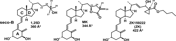
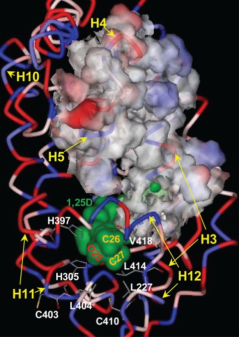
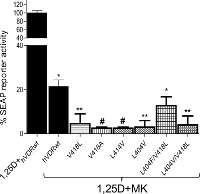
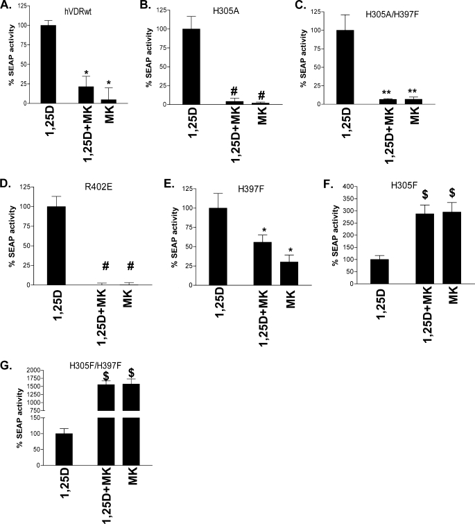
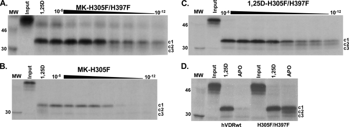
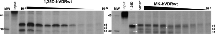
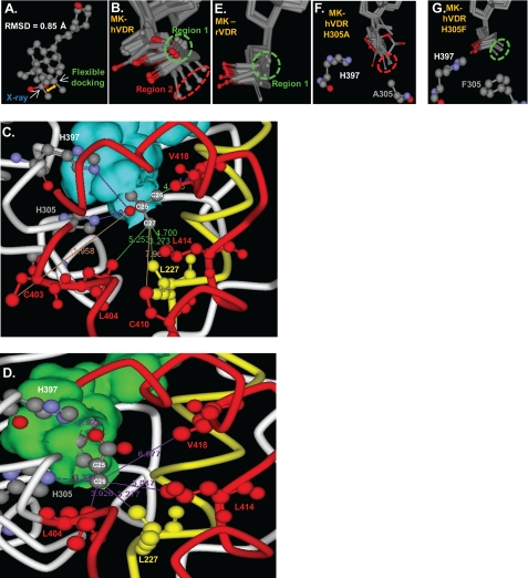
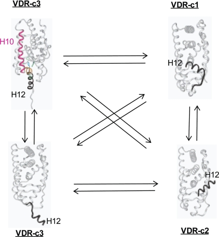
(23S)-25-Dehydro-1alpha(OH)-vitamin D(3)-26,23-lactone (MK) is an antagonist of the 1alpha,25(OH)(2)-vitamin D(3) (1,25D)/human nuclear vitamin D receptor (hVDR) transcription initiation complex, where the activation helix (i.e. helix-12) is closed. To study the mode of antagonism of MK an hVDR mutant library was designed to alter the free molecular volume in the region of the hVDR ligand binding pocket occupied by the ligand side-chain atoms (i.e. proximal to helix-12). The 1,25D-hVDR structure-function studies demonstrate that 1) van der Waals contacts between helix-12 residues Leu-414 and Val-418 and 1,25D enhance the stability of the closed helix-12 conformer and 2) removal of the side-chain H-bonds to His-305(F) and/or His-397(F) have no effect on 1,25D transactivation, even though they reduce the binding affinity of 1,25D. The MK structure-function results demonstrate that the His-305, Leu-404, Leu-414, and Val-418 mutations, which increase the free volume of the hVDR ligand binding pocket, significantly enhance MK antagonist potency. Surprisingly, the H305F and H305F/H397F mutations turn MK into a VDR superagonist (EC(50) approximately 0.05 nm) but do not concomitantly alter MK binding affinity. Molecular modeling studies demonstrate that MK antagonism stems from its side chain energetically preferring a pose in the VDR ligand binding pocket where its terminal C26-methylene atom is far removed from helix-12. MK superagonism results from an energetically favored increase in interaction between Leu-404/Val-418 and C26, resulting in an increase in the stability and population of the closed, helix-12 conformer. Finally, the results/model generated, coupled with application of a VDR ensemble allosterics model, provide an understanding for the species specificity of MK.


Figures

FIGURE 1.
FIGURE 1.
The chemistry of 1,25D, MK, and ZK. The chemical structures of 1α,25(OH)2-vitamin D3 (1,25D, C27H44O3) and the genomic antagonists of 1,25D-hVDR-mediated transactivation, MK (C27H38O4 (TEI-9647, Teijin Pharma)), and ZK (C32H48O5 (ZK159222, Schering)). The A, seco-B, C, and D rings of 1,25D are labeled, as are the side-chain carbon atoms C23, C24, C25 (arrow), C26, and C27. The molecular volumes of 1,25D, MK, and ZK were determined using DS2.0 (see “Experimental Procedures”).
FIGURE 2.
FIGURE 2.
The 1,25D-hVDR complex. The hVDR (LBD, aa 120–427, Δ165–215) is rendered to depict its ribbon structure and the relative hydrophobicity of its amino acid R-groups (blue, hydrophobic; red, polar, charged; pink-white, polar, uncharged). 1,25D is surface-rendered to illustrate its van der Waals (vdW) surface (green) and the location of the hVDR LBP. The position of the 1,25D C25 hydroxyl group and the C26/C27 side-chain methyl groups are labeled. The methyl groups lie proximal to Leu-227 helix-3 (H3), Leu-404 (H11), Leu-414 (H12), and Val-418 (H12), which form the hVDR LBP helix-3/helix-12 interface. The 25-OH group of 1,25D forms H-bonds with His-305 and His-397. The His-305 and Leu-404 residues form the bottom of the hVDR LBP. The upper surface of the nuclear co-activator (NCoA) binding site is highlighted by rendering the surface of amino acids 233 (H3) through 264 (H5) visible. The bottom of the NCoA surface is formed by helix-12 (H12). Helices 10 (H10) and 11 (H11) are labeled for reference.
FIGURE 3.
FIGURE 3.
Helix-3/helix-12 interface mutations increase the MK antagonist effect. CV1 cells were co-transfected with the osteocalcin VDR element-driven SEAP reporter and hVDRwt, L404V, L414V, V418A, V418L, L404F/V418L, and L404V/V418L plasmid constructs. The effective concentration (EC50, half-maximal activity) of 1,25D was measured in all the hVDR constructs, and a summary is provided in Table 1. The figure illustrates the % 1,25D SEAP activity measured when the cells were dosed with an EC50 concentration of 1,25D and a 10-fold excess of MK (n = 3). The values are expressed relative to the activity observed for 1,25D alone in each mutant, normalized to 100%. See supplemental Fig. 1 for representative 1,25D dose-response curves for these mutants. The ligand concentrations used are as follows: hVDRwt (1,25D = 1 nm; MK = 10 nm), L404V (1,25D = 40 nm; MK = 400 nm), K414V (1,25D = 25 nm; MK = 250 nm), V418A (1,25D = 75 nm; MK = 750 nm), V418L (1,25D = 1 nm; MK = 10 nm), L404V/V418L (1,25D = 500 nm; MK = 5 mm), L404V/L414V (1,25D = 75 nm; MK = 750 nm), and L404F/V418L (1,25D = 10 nm; MK = 100 nm). *, measured reporter activity is significantly reduced when compared with the 1,25D-induced SEAP-reporter activity in the given construct (p < 0.01; n ≥ 3, ±S.E.). **, SEAP-reporter activity is significantly reduced when compared with both the 1,25D-hVDRwt and 1,25D+MK·hVDRwt experiments (p < 0.05, ±S.E.; #, p < 0.01, ±S.E.).
FIGURE 4.
FIGURE 4.
Switching of MK into a complete antagonist/superagonist. CV1 cells were co-transfected with the osteocalcin VDR element-driven SEAP reporter and hVDRwt, H305A, H305A/H397F, H305F, H397F, or H305F/H397F plasmid constructs. A, compares the agonist potential of the EC50 dose of 1,25D alone (see Table 2), in the presence of a 10-fold excess of MK (1,25D + MK) and 10× MK alone in hVDRwt (n = 20). B–G depict the results obtained when CV1 cells were transfected with H305A, H305A/H397F, R402E, H397F, H305F, and H305F/H397F mutant constructs (n ≥ 3). The ligand concentrations were as follows: hVDRwt (1,25D = 1 nm; MK = 10 nm), H305A (1,25D = 30 nm; MK = 300 nm), H305A/H397F (1,25D = 30 nm; MK = 300 nm), R402E (1,25D = 10 nm; MK = 100 nm), H305F (1,25D = 1 nm; MK = 10 nm), H305F/H397F (1,25D = 1 nm; MK = 10 nm), and H397F (1,25D = 10 nm; MK = 100 nm). *, measured reporter activity is significantly reduced when compared with the 1,25D-induced SEAP-reporter activity in the given construct (p < 0.01; n ≥ 3, ±S.E.). **, SEAP-reporter activity is significantly reduced when compared with both the 1,25D-hVDRwt and 1,25D+MK·hVDRwt experiments (p < 0.05, ±S.E.; #, p < 0.01, ±S.E.). $, 1,25D + MK or MK agonist response is significantly better than the 1,25D SEAP-reporter induction in the same construct (p < 0.01, ±S.E.). Representative 1,25D and MK dose-response curves in these mutants are presented in supplemental Figs. 1 and 2.
FIGURE 5.
FIGURE 5.
1,25D and MK stabilize the hVDR closed conformation (hVDR-c1) in the H305F and H305F/H397F constructs. A–C depict the H305F/H397F and H305F trypsin fragments stabilized by MK and 1,25D (15% SDS-PAGE gels). In A and B the lane labeled 1,25D shows the PSA footprint stabilized by 10−5 m. Each gel represents an MK dose curve from 10 μm to 1 pm. Within the lanes in all panels, the hVDR-c1 (∼34 kDa, aa 174–427), -c2 (∼32 kDa, aa 174–413), and -c3 (∼30 kDa, aa 174–402) are labeled (20). D, 15% SDS-PAGE PSA footprint for apo/holo-hVDRwt and H305F/H397F. The concentration of 1,25D used in the holo-hVDR experiments was 10−5 m. For the MK-H305A, H397F, and H305A/H397F PSA footprint see supplemental Fig. 4.
FIGURE 6.
FIGURE 6.
Evidence MK forms covalent adducts with hVDRwt. Representative 12.5% SDS-PAGE gels showing the 1,25D (10 μm to 1 pm) and MK (1 μm to 1 nm) dose PSA results in hVDRwt, and the lane labeled 1,25D shows the PSA footprint stabilized by 10−5 m. The hVDR-c1 (∼34 kDa, aa 174–427), -c2 (∼32 kDa, aa 174–413), and -c3 (∼30 kDa, aa 174–402) are labeled (20). In the MK·hVDRwt gel (right panel) white arrows highlight what are concluded to be covalent adducts of c1, c2, and c3 formed by MK based on molecular weight shifts (see “Experimental Procedures”).
FIGURE 7.
FIGURE 7.
1,25D/MK·hVDR flexible docking results. A, superimposition of the 1,25D molecules extracted from the x-ray complex (25) and the lowest energy (ΔGbinding) flexible docking complex. The all atom root mean square deviation of 1,25D was 0.850 Å. Superimposition of the VDR Cα atoms of the same two complexes produced a root mean square deviation of 0.618 Å; all VDR backbone atoms were 0.635 Å; all VDR side-chain atoms were 0.956 Å. B, molecular overlay of the top 10 MK·hVDR LBP flexible docking complexes, depicting the two distinct MK side-chain conformational isomers observed in these complexes (Region 1, green dashed circle; Region 2, red dashed circle). C, the intermolecular interactions made between the 25-OH group and the C26 and C27 atoms of 1,25D observed in the lowest energy flexible docking complex (ΔGbinding). The VDR helix 11/helix-12 residues (Cys-403, Leu-404, Cys-410, Leu-414, and Val-418) are shown in red, and the helix-3, Leu-227 residue is shown in yellow. The same color scheme is used in panel D. The closest van der Waals (vdW) distance between Leu-227 (3.27 Å), Cys-403 (11.96 Å), Leu-404 (5.25 Å), Cys-410 (7.96 Å), Leu-414 (4.70 Å), Val-418 (4.03 Å), and the C26 or C27 terminal methyl groups of the 1,25D side-chain atoms are indicated by solid lines. The overlay of the top 10 1,25D-hVDRwt flexible docking complexes is presented in supplemental Fig. 5. D, the MK·hVDR LBP Region 2 complex and the intermolecular vdW interactions formed between the lactone C26 atom (Fig. 1) and Leu-227 (3.28 Å), His-305 (3.32 Å), His-397 (2.93 Å, to lactone ring oxygen), Leu-404 (3.93 Å), Leu-414 (5.05 Å), and Val-418 (6.98 Å) residues. These contacts are highlighted by purple lines. Importantly, the C26 atom of MK (Fig. 1) lies perpendicular to the sp2-hybrid imidazole nitrogen atom of His-305 in this complex, a good molecular geometry for formation of a covalent adduct (supplemental Fig. 6). E, the top 10 MK rodent VDR (rVDR) LBP flexible docking complexes indicate an enhanced selectivity for the MK side chain to localize in Region 1. To view the vdW interactions formed between the lactone of MK and the two His residues in the rVDR complex see supplemental Fig. 7. F, overlay of the top 10 H305A LBP flexible docking complexes. In all of these complexes the MK side chain occupies Region 2 of LBP, indicated by the red dashed circle. G, overlay of the top 10 MK-H305F LBP flexible docking complexes, indicating the C26 atom of MK that exclusively occupies Region 1 (green circle).
FIGURE 8.
FIGURE 8.
The VDR helix-12 conformational ensemble. Helix-12 of the VDR molecule is intrinsically disordered in the absence of ligand. In this figure the dynamic conformational heterogeneity of helix-12 of the hVDR is projected using NR x-ray data (see Protein Data Bank). It is observed that in hVDR-c1, helix-12 (black ribbon, labeled H12) is in the closed, active conformation (i.e. identical to its position in Fig. 2). It is proposed that, in the hVDR-c2 conformational isomer, helix-12 (black ribbon, labeled H12) is bound to the NCoA surface (Fig. 2) and is known to be induced by the antagonist analog ZK (Fig. 1) (5). It is proposed that in hVDR-c3, helix-12 (black ribbon, labeled H12) takes on an opened conformation, where the ligand binding pocket is accessible to ligand (bottom, hVDR-c3 structure) or is occupied by helix-11 residues (orange ribbon in the top hVDR-c3 structure), thereby exposing the C-terminal helix-11 Arg-402 residue (20).

Similar articles

Cited by

References

    1. Sutton A. L., MacDonald P. N. (2003) Mol. Endocrinol. 17, 777–791 - PubMed
    1. Cheskis B., Freedman L. P. (1994) Mol. Cell. Biol. 14, 3329–3338 - PMC - PubMed
    1. Barletta F., Freedman L. P., Christakos S. (2002) Mol. Endocrinol. 16, 301–314 - PubMed
    1. Rachez C., Freedman L. P. (2000) Gene 246, 9–21 - PubMed
    1. Tocchini-Valentini G., Rochel N., Wurtz J. M., Moras D. (2004) J. Med. Chem. 47, 1956–1961 - PubMed

MeSH terms

LinkOut - more resources