. 2009 Dec 11;284(50):35079-91.
doi: 10.1074/jbc.M109.040774. Epub 2009 Sep 29.

RNA helicase Prp43 and its co-factor Pfa1 promote 20 to 18 S rRNA processing catalyzed by the endonuclease Nob1

Affiliations

RNA helicase Prp43 and its co-factor Pfa1 promote 20 to 18 S rRNA processing catalyzed by the endonuclease Nob1

Brigitte Pertschy et al. J Biol Chem. .

Abstract

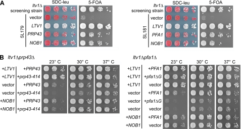
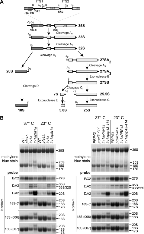
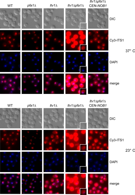
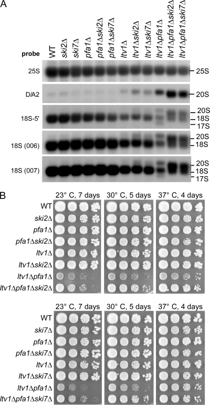
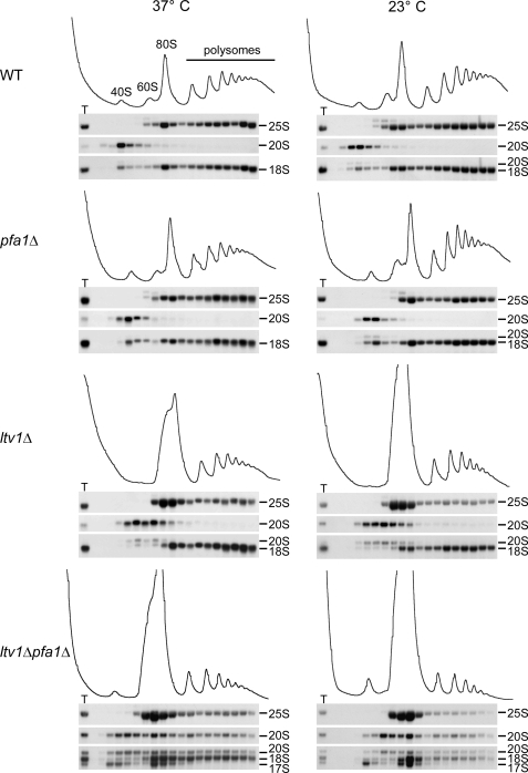
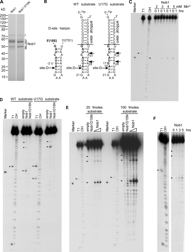
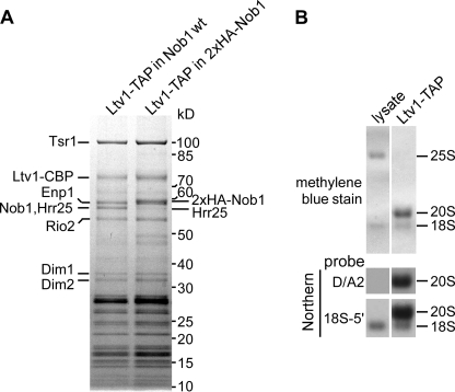
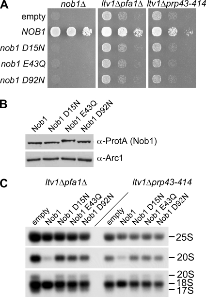
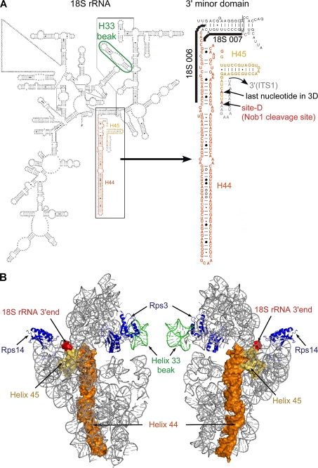
Many RNA nucleases and helicases participate in ribosome biogenesis, but how they cooperate with each other is largely unknown. Here we report that in vivo cleavage of the yeast pre-rRNA at site D, the 3'-end of the 18 S rRNA, requires functional interactions between PIN (PilT N terminus) domain protein Nob1 and the DEAH box RNA helicase Prp43. Nob1 showed specific cleavage on a D-site substrate analogue in vitro, which was abolished by mutations in the Nob1 PIN domain or the RNA substrate. Genetic analyses linked Nob1 to the late pre-40 S-associated factor Ltv1, the RNA helicase Prp43, and its cofactor Pfa1. In strains lacking Ltv1, mutation of Prp43 or Pfa1 led to a striking accumulation of 20 S pre-rRNA in the cytoplasm due to inhibition of site D cleavage. This phenotype was suppressed by increased dosage of wild-type Nob1 but not by Nob1 variants mutated in the catalytic site. In ltv1/pfa1 mutants the 20 S pre-rRNA was susceptible to 3' to 5' degradation by the cytoplasmic exosome. This degraded into the 3' region of the 18 S rRNA, strongly indicating that the preribosomes are structurally defective.


Figures

FIGURE 1.
FIGURE 1.
Genetic network between LTV1, PRP43, PFA1, and NOB1. A, identification of PRP43, PFA1, and NOB1 in a synthetic lethal screen with an ltv1 deletion strain. Mutants SL179 and SL181 isolated in the synthetic lethal (sl) screen were transformed with plasmids carrying the indicated genes and spotted in serial 10-fold dilution steps onto SDC−leu and SDC + 5-fluoroorotic acid (5-FOA) plates. As control, the non-mutagenized screening strain (ltv1Δ, pHT4467-CENΔ-LTV1) was spotted. Plates were incubated at 30 °C for 4 days. Red colony color on SDC−leu and slow growth on 5-fluoroorotic acid plates indicate a synthetic growth defect. Red/white sectoring on SDC−leu and growth on 5-fluoroorotic acid indicate complementation of the synthetic enhancement phenotype. B, temperature dependence of genetic interactions between LTV1 and PRP43 and between LTV1 and PFA1. The LTV1/PFA1 and LTV1/PRP43 shuffle strains were transformed with plasmids that carry the indicated wild-type and mutant alleles. pfa1ΔG is a PFA1 allele lacking the G-patch. After 5-fluoroorotic acid shuffling, cells were spotted in 10-fold serial dilution steps onto SDC−leu−trp plates to select for the transformed plasmids and incubated at 23, 30, and 37 °C for 4 days.
FIGURE 2.
FIGURE 2.
The interaction between Prp43 and Pfa1 is independent of the G-patch and resistant to increased salt concentrations. A, Pfa1-TAP was purified from yeast cells using increasing salt concentrations, ranging from 100 to 500 mm NaCl. Samples were analyzed by SDS-PAGE and Coomassie staining. The indicated proteins were identified by mass spectrometry. Note that at higher salt concentrations, only Pfa1 (full-length and degradation products) and Prp43 were recovered, indicating that Pfa1 and Prp43 directly interact in vivo. Pfa1-CBP, Pfa1 with the C-terminal calmodulin-binding protein tag. B, Pfa1- and Pfa1ΔG-TAP purifications. Note that the C-terminally truncated version of Pfa1 co-purified amounts of Prp43 similar to those provided by the full-length protein. All bands appearing in only one of the two purifications could be identified by mass spectrometry as N-terminal degradation products of Pfa1 that differed in size due to the different C termini of the bait proteins.
FIGURE 3.
FIGURE 3.
Ltv1, Pfa1, and Prp43 are required for 20 to 18 S rRNA processing. A, simplified rRNA processing pathway in yeast. Only the major pathway for generation of the 5′-end of the 5.8 S rRNA is shown. The rRNA cleavage sites and the binding sites of the probes used for Northern blotting are indicated. ITS1 and ITS2, internal transcribed spacers 1 and 2. In the course of pre-rRNA processing, the 35 S pre-rRNA undergoes a series of endonucleolytic processing events at sites A0, A1, and A2 that lead to the separation into the 20 S and 27 S A2 pre-rRNAs. Endo- and exonucleolytic processing steps of the 27 S A2 pre-rRNA finally yield the mature 25 and 5.8 S rRNAs contained in 60 S subunits. In the cytoplasm, the final processing step in 40 S maturation takes place when the 20 S pre-rRNA is converted into the 18 S rRNA by endonucleolytic cleavage at processing site D. B, rRNA steady state levels in ltv1Δ pfa1Δ and ltv1Δ prp43–414 mutants. Cells were either grown at 37 °C to an A600 of 0.8 (37 °C) or grown at 37 °C to an A600 of 0.1, transferred to 23 °C, and further grown to an A600 of 0.8 (three cell divisions). RNA was isolated, separated by agarose gel electrophoresis, and transferred to a nylon membrane that was stained with methylene blue. rRNA processing intermediates were detected by Northern blotting using the indicated probes. WT, wild type.
FIGURE 4.
FIGURE 4.
20 S pre-rRNA accumulates in the cytoplasm in ltv1Δ pfa1Δ mutants. ltv1Δ pfa1Δ cells transformed with empty plasmids or plasmids carrying LTV1, PFA1, or NOB1 wild-type alleles were either grown at 37 °C to an A600 of 0.7–1 (37 °C) or grown to an A600 of 0.3, and shifted to 23 °C for 3 h (23 °C). Cells were fixed with formaldehyde, spheroplasted, and subjected to fluorescence in situ hybridization using a Cy3-labeled probe complementary to a sequence in the D/A2 segment of ITS1. To visualize the nucleoplasm, cells were stained with 4′,6-diamidino-2-phenylindole (DAPI). Due to the strong signal in the ltv1Δ pfa1Δ cells, also single cells with a shorter exposure of the Cy3 signal are shown. WT, wild type.
FIGURE 5.
FIGURE 5.
17 S RNA is generated via an 3′–5′ exonucleolytic attack by the exosome. A, the abnormal 17 S RNA species is not formed when SKI2 or SKI7 is deleted. Cells were grown at 30 °C to an A600 of 0.8. RNA was isolated and analyzed by Northern blotting using the indicated probes. B, ltv1Δ pfa1Δ mutants grow faster when also SKI2 or SKI7 is deleted. Growth of ski2Δ (top) and ski7Δ (bottom) mutants is shown. Cells were spotted in 10-fold serial dilution steps onto YPD plates and incubated at the indicated temperatures for the indicated time periods. WT, wild type.
FIGURE 6.
FIGURE 6.
17 S RNA co-sediments only with 40 and 80 S, whereas 20 S pre-rRNA is also found in polysomes. Cells were either grown at 37 °C to an A600 of 0.7–1 (37 °C) or grown to an A600 of 0.1, shifted to 23 °C, and incubated to an A600 of 0.7–1 (23 °C). Cell extracts were loaded onto 7–50% sucrose gradients and fractionated. RNA was extracted from each fraction and analyzed by Northern blotting using the probes 25 S, D/A2 (to detect 20 S pre-rRNA), and 5′-18 S (to detect 20, 18, and 17 S rRNAs). T, RNA preparation of the total extract. WT, wild type.
FIGURE 7.
FIGURE 7.
Nob1 has D-site-specific endonuclease activity in vitro. A, purified wild-type Nob1 and mutant Nob1-D15N used in the in vitro nuclease assay. The proteins were affinity-purified from yeast and analyzed by SDS-PAGE and Coomassie staining. All bands labeled with Nob1 were identified by mass spectrometry. Hsp70 contaminants are indicated by stars. B, model of the cleavage site D within the 20 S pre-rRNA. Left sequence, authentic D-site hairpin ranging from 5′ (part of mature 18 S rRNA; shown in boldface type) to 3′ (part of ITS1, shown in normal type) (53); middle and right sequences, RNA model substrates with engineered wild-type (middle) and mutated cleavage site D (U17G mutation) (right). Both RNA substrates carry an additional stem sequence attached to the D-site hairpin. Alternative cleavage sites at positions A28 and A31 are indicated. C–F, Nob1 in vitro cleavage assays. Marker, 18-nucleotide RNA resembling the expected D-site cleavage product, ranging from nucleotide 1 to 18 of the used WT substrate. T1, substrate digested with T1 nuclease, which cleaves after single-stranded G residues (G1 and G21, as well as G17 in the U17G substrate). OH, partial alkaline hydrolysis of the RNA substrate. Due to the generation of 3′-phosphates upon hydrolysis by T1 nuclease or OH, the products run slightly faster (∼1.5 nucleotides) than cleavage products with 3′-OH ends. 3′-OH ends are found in the marker and in cleavage products predicted to be generated by PIN endonucleases, as shown for the exosome subunit Rrp44 (14). Note that the cleavage product of Nob1 runs at a similar height as the A18 oligonucleotide, indicating that cleavage occurs after A18, corresponding to the D-site. Small variations in cleavage efficiencies in the assays shown are probably due to the use of different protein and/or RNA substrate preparations. C, manganese titration. 5′-[32P]-end-labeled RNA substrate was incubated with purified wild-type Nob1 at 30 °C for 0 or 1 h in the presence of increasing concentrations of manganese as indicated. ∼5 pmol of Nob1 and 10 fmol of substrate were used (500-fold molar excess). The arrows indicate the G21 cleavage product generated by T1 nuclease and the A18 cleavage product generated by Nob1. D, cleavage does not occur when either the catalytic site of Nob1 (Nob1-D15N) or the sequence of the substrate (U17G) is mutated. 10 fmol of 5′-32P-end-labeled RNA substrates (shown in B) were incubated without protein (empty) or with purified wild-type Nob1 or mutant Nob1-D15N (5 pmol each) at 30 °C for 1 h. The arrows indicate the G17 and G21 cleavage products generated by T1 nuclease and the A18, A28, and A31 cleavage products generated by Nob1. E, dependence of the cleavage reaction on enzyme and substrate concentration. 20 (left) and 100 (right) fmol of RNA substrate were incubated with 1.25, 2.5, or 5 pmol of Nob1 protein for 1 h at 30 °C. In the case of the Nob1-D15N protein, 5 pmol were used. The arrows indicate the G21 cleavage product generated by T1 nuclease and the A18, A28, and A31 cleavage products generated by Nob1. F, time course of Nob1 cleavage. 20 fmol of substrate were incubated with 10 pmol of Nob1 at 30 °C for the indicated time periods. The arrows indicate the G21 cleavage product generated by T1 nuclease and the A18, A28, and A31 cleavage products generated by Nob1. WT, wild type.
FIGURE 8.
FIGURE 8.
pre-40 S particles purified via Ltv1-TAP contain Nob1 and unprocessed 20 S pre-rRNA. A, Nob1 is stably associated with the pre-40 S particle affinity-purified via Ltv1-TAP. The Ltv1-TAP preparation was analyzed by SDS-PAGE and Coomassie staining. Bands identified by mass spectrometry are indicated. Nob1 and Hrr25 co-migrate as one band. To estimate the proportion of Nob1 in this band, we performed another purification where the size of Nob1 was increased by fusion to an N-terminal hemagglutinin tag. B, the main RNA species in the Ltv1-TAP purification is 20 S pre-rRNA. RNA was extracted from the cell lysate and the pre-40 S subunit purified via Ltv1-TAP, separated on an agarose gel and transferred to a nylon membrane that was stained with methylene blue. The 20 and 18 S rRNAs were detected by Northern blotting with specific probes (see Fig. 3A).
FIGURE 9.
FIGURE 9.
Suppression of the rRNA processing defects in ltv1Δ pfa1Δ and ltv1Δ prp43–414 mutants requires the endonuclease activity of Nob1. A, to test whether the nob1 mutant alleles are functional in vivo, plasmids expressing Nob1 mutants that were N-terminally tagged with ProtA-FLAG were transformed into a NOB1 shuffle strain, and growth was tested by spotting cells in 10-fold dilution steps onto plates containing 5-fluoroorotic acid (5-FOA) and incubated at 30 °C for 2 days. Growth indicates complementation of the nob1 deletion. To test for suppression, plasmids were transformed into the double mutants ltv1Δ pfa1Δ and ltv1Δ prp43–414, and transformants were spotted onto SDC−leu plates. Plates were incubated at 30 °C for 4 days. B, expression of the indicated Nob1 mutant proteins was tested by analyzing whole cell lysates from a NOB1 shuffle strain transformed with the respective plasmids and Western blotting against the ProtA tag. The Western blot using an antibody against Arc1 serves as a loading control. Note that the electrophoretic mobility of the mutant variants is slightly changed, which could be due to the removed negative charge. C, Northern blotting of ltv1Δ pfa1Δ and ltv1Δ prp43–414 strains transformed with plasmids carrying the indicated ProtA-FLAG-tagged Nob1 variants. Cells were grown at 30 °C to an A600 of 0.8. RNA was isolated and analyzed by Northern blotting using the 25 S probe to detect 25 S rRNA, the D/A2 probe to detect 20 S pre-rRNA, and the 18 S 5′ probe to detect 20, 18, and 17 S rRNAs.
FIGURE 10.
FIGURE 10.
Structure of the 3′-end of the 18 S rRNA. A, left, modeled secondary structure of the 18 S rRNA according to Refs. and 55). Helix 33 is encircled in green. The box indicates the 3′ minor domain, consisting of helices 44 (orange) and 45 (yellow). Right, enlarged view of the 3′ minor domain. Binding sites of oligonucleotides 18 S 006 and 007 are indicated. The last nucleotide in the three-dimensional model shown in B and the actual 3′-end of the 18 S rRNA are displayed in red. The sequence of ITS1 within the predicted D-site hairpin (53) was added manually (gray). B, modeled structure of the yeast 40 S subunit, shown from the solvent (left) and intersubunit (right) sides. Helices 44 (orange) and 45 (yellow) corresponding to the last ∼160 nucleotides at the 3′-end of the 18 S rRNA are shown as a surface representation. The 3′-end of the 18 S rRNA is displayed in red, and helix 33 is shown in green. Of the ribosomal proteins, only Rps3 and Rps14 are shown (blue). The displayed images were generated in PyMOL (DeLano Scientific).

References

    1. Fatica A., Tollervey D. (2002) Curr. Opin. Cell Biol. 14, 313–318 - PubMed
    1. Fromont-Racine M., Senger B., Saveanu C., Fasiolo F. (2003) Gene 313, 17–42 - PubMed
    1. Granneman S., Baserga S. J. (2004) Exp. Cell Res. 296, 43–50 - PubMed
    1. Henras A. K., Soudet J., Gérus M., Lebaron S., Caizergues-Ferrer M., Mougin A., Henry Y. (2008) Cell Mol. Life Sci. 65, 2334–2359 - PMC - PubMed
    1. Tschochner H., Hurt E. (2003) Trends Cell Biol. 13, 255–263 - PubMed

Publication types

MeSH terms