. 2009 Nov 20;284(47):32642-50.
doi: 10.1074/jbc.M109.046573. Epub 2009 Sep 30.

Identification of a new exosite involved in catalytic turnover by the streptokinase-plasmin activator complex during human plasminogen activation

Affiliations

Identification of a new exosite involved in catalytic turnover by the streptokinase-plasmin activator complex during human plasminogen activation

Rachna Aneja et al. J Biol Chem. .

Abstract

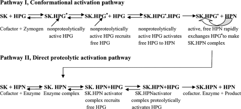
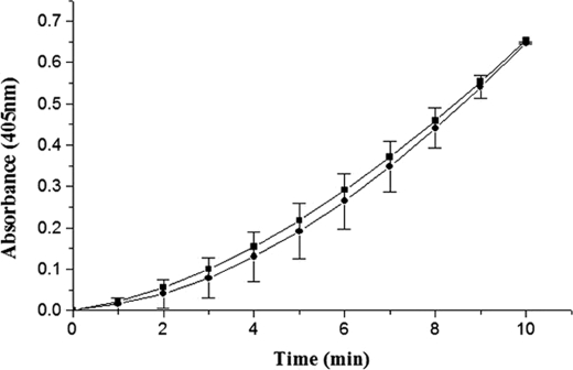
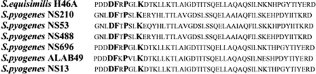
With the goal of identifying hitherto unknown surface exosites of streptokinase involved in substrate human plasminogen recognition and catalytic turnover, synthetic peptides encompassing the 170 loop (CQFTPLNPDDDFRPGLKDTKLLC) in the beta-domain were tested for selective inhibition of substrate human plasminogen activation by the streptokinase-plasmin activator complex. Although a disulfide-constrained peptide exhibited strong inhibition, a linear peptide with the same sequence, or a disulfide-constrained variant with a single lysine to alanine mutation showed significantly reduced capabilities of inhibition. Alanine-scanning mutagenesis of the 170 loop of the beta-domain of streptokinase was then performed to elucidate its importance in streptokinase-mediated plasminogen activation. Some of the 170 loop mutants showed a remarkable decline in k(cat) without any alteration in apparent substrate affinity (K(m)) as compared with wild-type streptokinase and identified the importance of Lys(180) as well as Pro(177) in the functioning of this loop. Remarkably, these mutants were able to generate amidolytic activity and non-proteolytic activation in "partner" plasminogen as wild-type streptokinase. Moreover, cofactor activities of the 170 loop mutants, pre-complexed with plasmin, against microplasminogen as the substrate showed a similar pattern of decline in k(cat) as that observed in the case of full-length plasminogen, with no concomitant change in K(m). These results strongly suggest that the 170 loop of the beta-domain of streptokinase is important for catalysis by the streptokinase-plasmin(ogen) activator complex, particularly in catalytic processing/turnover of substrate, although it does not seem to contribute significantly toward enzyme-substrate affinity per se.


Figures

FIGURE 1.
FIGURE 1.
Currently understood mechanism of plasminogen activation by streptokinase. SK forms an equimolar complex with HPG (Pathway I) that leads to the generation of an active center in the complex (SK·HPG*) without cleavage of scissile peptide bond in the partner HPG. This “virgin complex” then recruits free HPG as substrate and converts the latter into HPN. The HPN so formed rapidly exchanges with the HPG* from SK·HPG* complex due to its multifold higher affinity for SK in comparison to HPG. Alternatively, the SK can directly combine with the HPN to make SK·HPN activator complex (Pathway II). The activator complex then catalytically acts on substrate HPG molecules and converts them into HPN.
FIGURE 2.
FIGURE 2.
Selective inhibition of substrate HPG activation ability of wtSK·HPN activator complex by 170 loop peptide mimotopes. Aliquots of preformed SK·HPN complex (5 nm) were added to HPG (0.05 μm) in assay buffer containing various concentrations of the 170 loop sequence peptides (0–500 μm). Residual percent activity in the presence of either linear, unconstrained 170 loop peptide (open circles), wild-type cyclic 170 loop peptide (solid squares), linear K180A peptide (solid circles), or the disulfide constrained cyclic K180A peptide (solid diamonds), was calculated from the rates of plasmin generated in the presence of peptide compared with that of activation reaction (taken as 100%) where no peptide was added (see “Experimental Procedures” for details). The control reactions where either plasmin inhibition (in the absence of SK) was tested in the presence of peptides are also depicted (upward triangles), or where possible inhibition of SK·HPN activator complex in the absence of substrate plasminogen (downward triangles) are also depicted.
FIGURE 3.
FIGURE 3.
Time course of single stage HPG activation assays by wtSK and 170 loop mutants. SK/various 170 loop mutants of SK (0.5 nm each) were added individually to microtiter plate wells containing HPG and Chromozym®PL, and the activator activities were measured spectrophotometrically at 405 nm as detailed under “Experimental Procedures.” The graph shows wild-type SK (solid squares), R176A (solid circles), P177A (upward triangles), K180A (inverted triangles), K180G (open circles), K180D (solid diamonds), and D181A (open squares).
FIGURE 4.
FIGURE 4.
Active site titration of HPG upon complexing with wtSK/K180A mutant using the active site acylating agent, NPGB. The figure shows progress curves of NPGB hydrolysis as followed by p-nitrophenol liberation by MetAP-treated wtSK (open upward triangles), and MetAP-treated K180A (solid diamonds) complexes with HPG, and a control reaction (solid squares) where no activator was taken (see “Experimental Procedures” for details).
FIGURE 5.
FIGURE 5.
Time course of the amidolytic activity generation in R561A μPG by wtSK and K180A mutant. Equimolar SK·R561A μPG mutant and K180A·R561A μPG mutant complexes were made, and catalytic amounts were added to the assay buffer containing 1 mm chromogenic substrate Chromozym®PL, 50 mm Tris, pH 7.4, 100 mm NaCl, and amidolytic activity generated was represented as wild-type SK (solid squares) or K180A mutant (solid circles). Assays were performed at 37 °C with 10 nm SK and μPG complex as described under “Experimental Procedures.”
FIGURE 6.
FIGURE 6.
Multiple alignment of primary sequence of β-domain depicting the 170 loop amino acid residues of streptokinases secreted from S. equisimilis and three representative allelic variant clusters of Streptococcus pyogenes, namely strains NS210, NS53, NS488, NS696, ALAB49, and NS13 (see Ref. 34). Note that residues (all given in single-letter code) highlighted in bold type are conserved in 170 loop in all the streptokinase polypeptides obtained from the different sources.

References

    1. Lijnen H. R., Stassen J. M., Vanlinthout I., Fukao H., Okada K., Matsuo O., Collen D. (1991) Thromb. Haemost. 66, 468–473 - PubMed
    1. McClintock D. K., Bell P. H. (1971) Biochem. Biophys. Res. Commun. 43, 694–702 - PubMed
    1. Bajaj A. P., Castellino F. J. (1977) J. Biol. Chem. 252, 492–498 - PubMed
    1. Boxrud P. D., Verhamme I. M., Bock P. E. (2004) J. Biol. Chem. 279, 36633–36641 - PubMed
    1. Boxrud P. D., Bock P. E. (2004) J. Biol. Chem. 279, 36642–36649 - PubMed

Publication types

LinkOut - more resources