. 2009 Nov 27;284(48):33019-29.
doi: 10.1074/jbc.M109.034553. Epub 2009 Oct 1.

The miR200 family of microRNAs regulates WAVE3-dependent cancer cell invasion

Affiliations

The miR200 family of microRNAs regulates WAVE3-dependent cancer cell invasion

Khalid Sossey-Alaoui et al. J Biol Chem. .

Abstract

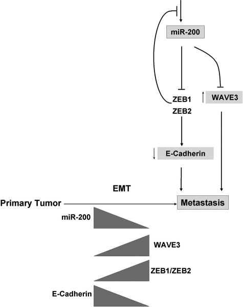
MicroRNAs are small non-coding RNAs that are directly involved in the regulation of gene expression by either translational repression or degradation of target mRNAs. Because of the high level of conservation of the target motifs, known as seed sequences, within the 3'-untranslated regions, a single microRNA can regulate numerous target genes simultaneously, making this class of RNAs a powerful regulator of gene expression. The miR200 family of microRNAs has recently been shown to regulate the process of epithelial to mesenchymal transition during tumor progression and metastasis. Here, we report that the expression of WAVE3, an actin cytoskeleton remodeling and metastasis promoter protein, is regulated by miR200 microRNAs. We show a clear inverse correlation between expression levels of WAVE3 and miR200 microRNAs in invasive versus non-invasive cancer cells. miR200 directly targets the 3'-untranslated regions of the WAVE3 mRNA and inhibits its expression. The miR200-mediated down-regulation of WAVE3 results in a significant reduction in the invasive phenotype of cancer cells, which is specific to the loss of WAVE3 expression. Re-expression of a miR200-resistant WAVE3 reverses miR200-mediated inhibition of cancer cell invasion. Loss of WAVE3 expression downstream of miR200 also results in a dramatic change in cell morphology resembling that of a mesenchymal to epithelial transition. In conclusion, a novel mechanism for the regulation of WAVE3 expression in cancer cells has been identified, which controls the invasive properties and morphology of cancer cells associated with their metastatic potential.


Figures

FIGURE 1.
FIGURE 1.
WAVE3 expression is elevated in cell lines with low levels of miR200 microRNAs. A, domain structure of the WAVE3 transcript showing the location of the seed sequence of the miR200 family of microRNAs within the 3′-UTR. B, schematic representation of the miR200b-200a-429 polysistronic cluster indicating the position of the putative transcription start site (TSS) and the polyadenylation signal. The locations of amplicons A–F are depicted with horizontal solid black bars. The preservation of the genomic structure and mRNA expression along the cluster was verified by genomic PCR (C) and RT-PCR (D), respectively. ORF, open reading frame; GAPDH, glyceraldehyde-3-phosphate dehydrogenase.
FIGURE 2.
FIGURE 2.
microRNA miR200b directly targets the 3′-UTR of WAVE3 and represses its expression. A, RT-PCR analysis of WAVE3 in untreated HEK293 cells or transfected with miR200b. WAVE3 expression levels were markedly reduced in the miR200b-transfected cells. WAVE2 and glyceraldehyde-3-phosphate dehydrogenase (GAPDH) were used as controls. B, firefly luciferase reporter plasmids, pmirGlo control, or pmirGlo containing the entire 3′-UTR of WAVE3 (W3–3UTR) were transiently transfected into HEK293 cells, and luciferase activities were measured after 48 h. C, HEK293 cells were transfected with luciferase reporter plasmids or co-transfected with luciferase reporter plasmids and synthetic miR200b mimic; luciferase activities were measured after 48 h.
FIGURE 3.
FIGURE 3.
Mutation of miR200 seed sequences in the WAVE3 3′-UTR abrogates miR200b effect. A and B, RT-PCR analysis of WAVE3 in MDA-MB-231 (A) and LNCaP (B) transiently transfected with either siRNA to WAVE3, synthetic miR200b, or control microRNA. C and D, MDA-MB-231, LNCaP, and HT29 cells were transfected with LR control or WAVE3–3′-UTR plasmids (C) or co-transfected with LR plasmids and synthetic miR200b precursor (D), and luciferase activities were measured after 48 h. E, MDA-MB-231, LNCaP, and HT29 cells were transfected with LR plasmids containing the WAVE3 3′-UTR with deleted miR200b seed sequences and co-transfected with or without synthetic miR200b precursor, and luciferase activities were measured after 48 h. For all luciferase activity assays, Renilla luciferase activity was used for normalization. The data are the mean ± S.D. of at least three independent transfections. GAPDH, glyceraldehyde-3-phosphate dehydrogenase.
FIGURE 4.
FIGURE 4.
miR200-mediated down-regulation of WAVE3 expression inhibits cancer cell invasion. Matrigel invasion assay of MDA-MB-231 (A and B), LNCaP (C and D), and HT29 (E and F) transfected with either siRNA to WAVE3 or miR200b precursor. Shown are representative bright field microscopy images of stained cells (A, C, and E) and quantification of invasive cells (B, D, and F). At least six different fields were counted from each experiment. The data are the mean ± S.D. of at least three independent assays.
FIGURE 5.
FIGURE 5.
Re-expression of miR200-resistant WAVE3 reverses miR200b-mediated inhibition of LNCaP cell invasion. A, immunoblotting (IB) with the indicated antisera of LNCaP cells overexpressing green fluorescent protein or green fluorescent protein-WAVE3 fusion protein and co-transfected with either control siWAVE3 of miR200b. NS, nonspecific band. B, quantification of invasive cells for the indicated treatment. At least six different fields were counted from each experiment. The data are the mean ± S.D. of at least three independent assays.
FIGURE 6.
FIGURE 6.
miR200 regulates WAVE3 and E-cadherin/ZEB1/ZEB2 through independent pathways during EMT. Western blot (A, C, and E) and RT-PCR (B, D, and F) analyses of WAVE3, E-cadherin, and vimentin in MDA-MB-231, LNCaP, and HT29, respectively, with the indicated transient treatments. Glyceraldehyde-3-phosphate dehydrogenase (GAPDH) and β-actin were used as internal controls for RT-PCR and Western blot analyses, respectively.
FIGURE 7.
FIGURE 7.
Mechanism of miR200-mediated regulation of WAVE3 during EMT. Tumor progression from the primary to the invasive/metastatic stage is associated with an increase in the expression levels of ZEB1 and ZEB2 inhibitors of transcription factors, leading to a significant decrease in the expression levels of miR200 microRNAs, which results in an increase in the expression levels of WAVE3 and a concomitant decrease in E-cadherin expression levels. Whereas down-regulation of E-cadherin leads to the loss of cell junctions, up-regulation of WAVE3 results in an increase in cell motility, both of which are required for the acquisition of the invasive phenotype. This coordinated regulation of both E-cadherin and WAVE3 by miR200 microRNAs is critical for tumor progression and metastasis.

References

    1. Berx G., Raspé E., Christofori G., Thiery J. P., Sleeman J. P. (2007) Clin. Exp. Metastasis 24, 587–597 - PubMed
    1. Burk U., Schubert J., Wellner U., Schmalhofer O., Vincan E., Spaderna S., Brabletz T. (2008) EMBO Rep. 9, 582–589 - PMC - PubMed
    1. Esquela-Kerscher A., Slack F. J. (2006) Nat. Rev. Cancer 6, 259–269 - PubMed
    1. Korpal M., Kang Y. (2008) RNA Biol. 5, 115–119 - PMC - PubMed
    1. Korpal M., Lee E. S., Hu G., Kang Y. (2008) J. Biol. Chem. 283, 14910–14914 - PMC - PubMed

Publication types

MeSH terms