Review
. 2009 Oct;32(10):1639-44.
doi: 10.1248/bpb.32.1639.

Oligovascular signaling in white matter stroke

Affiliations
Review

Oligovascular signaling in white matter stroke

Ken Arai et al. Biol Pharm Bull. 2009 Oct.

Abstract

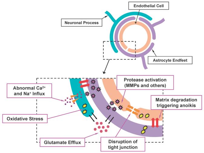
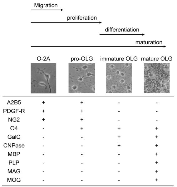
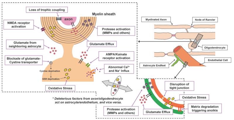
Stroke is one of the leading causes of death and disability in developed countries. Since protecting neurons alone is not sufficient for stroke therapy, research has shifted to the rescue of multiple cell types in the brain. In particular, attention has focused on the study of how cerebral blood vessels and brain cells communicate with each other. Recent findings suggest that cerebral endothelial cells may secrete trophic factors that nourish neighboring cells. Although data are strongest in terms of supporting endothelial-neuronal interactions, it is likely that similar interactions occur in white matter as well. In this mini-review, we summarize recent advances in the dissection of cell-cell interactions in white matter. We examine two key concepts. First, trophic interactions between vessels and oligodendrocytes (OLGs) and oligodendrocyte precursor cells (OPCs) play critical roles in white matter homeostasis. Second, cell-cell trophic coupling is disturbed under diseased conditions that incur oxidative stress. White matter pathophysiology is very important in stroke. A deeper understanding of the mechanisms of oligovascular signaling in normal and pathologic conditions may lead us to new therapeutic targets for stroke and other neurodegenerative diseases.


Figures

Fig. 1
Fig. 1
Schematic of the Neurovascular Unit under Stroke Conditions Neurons, astrocytes, cerebral endothelial cells, and extracellular matrix comprise the neurovascular unit. This concept emphasizes the functional aspects of cell–cell and cell–matrix signaling. Under normal conditions, these components work together to maintain brain function. After ischemia, neurovascular damage occurs by multiple deleterious pathways, resulting in loss of brain function. The reader is encouraged to seek more detailed reviews describing these events.
Fig. 2
Fig. 2
Characteristics of OLG Maturation OLG maturation is defined by the expression of specific cell-surface receptors, cell morphology, and proliferative/motility responses. O-2A cells are bipolar cells with high proliferative and motile activities. Pro-OLGs still divide but no longer show motility response. Immature OLGs are one of the two differenciated OLGs, but do not form myelin yet. The other differenciated OLG is mature OLGs, which express the myelin proteins to form the myelin sheath around axons. The reader is encouraged to seek more detailed reviews describing the full complexities of OLG maturation.
Fig. 3
Fig. 3
Schematic of the Adult White Matter under Stroke Conditions The main components of white matter are the neuronal axon, oligodendrocyte (myelin), astrocyte, and endothelium. Similar to gray matter, cell-cell interactions are important to maintain white matter function. Under ishemic conditions, several deleterious factors/cascades are activated. As in the neurovascular unit, several events occur due to ischemic stress. Glutamate efflux, oxidative stress, and proteinase activation eventually induce cell death. Importantly, deleterious factors secreted by one cell type may affect another cell type. For example, cerebral endothelial cells secrete MMPs after ischemic injury. These MMPs, in turn, damage the myelin sheath produced by oligodendrocytes. Moreover, under ischemic conditions, trophic support from astrocyte/endothelium to myelinated axons is disturbed by oxidative stress. The reader is encouraged to seek more detailed reviews describing the events in white matter ischemia.—,
Fig. 4
Fig. 4
Schematic of the Oligovascular Signaling in Adult White Matter In the oligovascular niche, oligogenesis and angiogenesis might occur to maintain white matter homeostasis. Oligodendrocyte precursor cells (OPCs) are thought to contribute to myelin maintenance and repair by generating new mature oligodendrocytes (OLGs). In addition, trophic coupling may also exist between cerebral endothelial cells and mature OLGs. Under diseased conditions such as stroke, multiple deleterious factors directly attack OLGs and OPCs, which are vulnerable to oxidative stress. Moreover, oxidative stress can disturb the trophic coupling between cerebral endothelial cells and OLGs/OPCs, resulting in further white matter damage.

Similar articles

Cited by

References

    1. Guo S, Lo EH. Stroke. 2009;40:S4–S7. - PMC - PubMed
    1. Lo EH, Dalkara T, Moskowitz MA. Nat. Rev. Neurosci. 2003;4:399–415. - PubMed
    1. del Zoppo GJ. N. Engl. J. Med. 2006;354:553–555. - PubMed
    1. Iadecola C. Nat. Rev. Neurosci. 2004;5:347–360. - PubMed
    1. Zlokovic BV. Neuron. 2008;57:178–201. - PubMed

Publication types