. 2009 Nov;11(11):1370-5.
doi: 10.1038/ncb1981. Epub 2009 Oct 4.

Transcriptional repression of p53 by parkin and impairment by mutations associated with autosomal recessive juvenile Parkinson's disease

Affiliations

Transcriptional repression of p53 by parkin and impairment by mutations associated with autosomal recessive juvenile Parkinson's disease

Cristine Alves da Costa et al. Nat Cell Biol. 2009 Nov.

Abstract

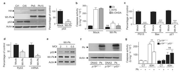
Mutations of the ubiquitin ligase parkin account for most autosomal recessive forms of juvenile Parkinson's disease (AR-JP). Several studies have suggested that parkin possesses DNA-binding and transcriptional activity. We report here that parkin is a p53 transcriptional repressor. First, parkin prevented 6-hydroxydopamine-induced caspase-3 activation in a p53-dependent manner. Concomitantly, parkin reduced p53 expression and activity, an effect abrogated by familial parkin mutations known to either abolish or preserve its ligase activity. ChIP experiments indicate that overexpressed and endogenous parkin interact physically with the p53 promoter and that pathogenic mutations abolish DNA binding to and promoter transactivation of p53. Parkin lowered p53 mRNA levels and repressed p53 promoter transactivation through its Ring1 domain. Conversely, parkin depletion enhanced p53 expression and mRNA levels in fibroblasts and mouse brains, and increased cellular p53 activity and promoter transactivation in cells. Finally, familial parkin missense and deletion mutations enhanced p53 expression in human brains affected by AR-JP. This study reveals a ubiquitin ligase-independent function of parkin in the control of transcription and a functional link between parkin and p53 that is altered by AR-JP mutations.


Figures

Figure 1
Figure 1
Protective effect of parkin is associated with modulation of p53 in TSM1 neuronal cell line. (a) Analysis of endogenous (Wt-Pk) and HA-tagged-parkin (HA), p53 and actin-like immunoreactivity in stably transfected TSM1 cells overexpressing either wild-type parkin (Pk) or empty vector (Ct) (numbers indicate distinct clones). The right panel shows the densitometric analysis of p53-like immunoreactivity (normalized to actin expression) expressed as percentage of control mock-transfected cells. Bars represent the mean ± s.e.m. of 3 independent experiments performed in duplicate (**P < 0.001). (b) Determination of caspase-3 activity in TSM1 neurons overexpressing empty vector (mock) and wild-type parkin (Wt-Pk) after treatment with staurosporine (STS, 1 μM) for 2 h or 6-hydroxydopamine (6OHDA, 0.2 mM) for 8 h. Bars represent the mean ± s.e.m. of 5 independent experiments performed in duplicate (*P <0.01 for Wt-Pk compare with Mock). (c, d) Analysis of p53 activity (PG13, Bax and p21), promoter transactivation (Pp53) and mRNA levels (mRNA) in Mock- and Wt-Pk-transfected cells (clone Pk15, see a). Values are normalized for transfection efficiency (β-galactosidase activity), expressed as percentage of mock-transfected cells (taken as 100) and are the mean ± s.e.m. of 4 (Pp53, mRNA and PG13) or 5 (Bax and p21) independent experiments performed in duplicate (*P <0.01, **P <0.001, ***P <0.0001). (e) p53 expression in lentiviral-infected primary cultured neurons overexpressing parkin. Primary cultured neurons were infected with 0.5, 1 and 2 MOI (multiplicity of infection) of Wt-Pk-lentiviral vector and then assayed for parkin and p53 expression four days after infection. (f, g) p19arf−/− and p19arf−/−/p53−/− fibroblasts were transiently transfected with empty pcDNA3 (DNA3) or parkin (Pk) cDNA. Twenty-four hours after transfection, cells were treated with 6-OHDA (6OH, 0.2 mM) for 8 h then expression of parkin (f) and caspase-3 activity (g) were monitored. Bars represent mean ± s.e.m. of 3 independent experiments performed in triplicate (**P <0.001; ns, not significant). Parkin and actin levels were assessed on separate gels as explained in Methods. Full scans of the blots in a, e and f are available in Supplementary Information, Fig.S3.
Figure 2
Figure 2
p53 pathway is upregulated in parkin-deficient fibroblasts and mouse brains. (a) Caspase-3 activity in 6-OHDA-treated (0.2 mM, 8 h) wild-type (Pk+ and parkin-deficient (Pk) fibroblasts. Bars represent the mean ± s.e.m. of 3 independent experiments performed in triplicate (*P <0.05; **P <0.01). (b) p53 and actin expression in Pk+ and Pk fibroblasts were determined by western blotting. The lower panel shows densitometric analysis of p53 (normalized by actin expression) and bars represent mean ± s.e.m. of 3 independent experiments performed in duplicate (**P <0.01). (c) Analysis of p53 activity (PG13), promoter transactivation (Pp53) and mRNA levels in Pk+ and Pk fibroblasts. Values normalized for transfection efficiency (β-galactosidase activity) are expressed as percentage of mock-transfected cells (taken as 100). Bars represent the mean ± s.e.m. of 3 independent experiments performed in duplicate (*P <0.05; **P <0.01). (d) p53 and actin immunoreactivity in brain homogenates derived from Pk+ and Pk mice were determined. The right panel shows densitometric analysis of p53 (normalized by actin levels) and bars represent the mean ± s.e.m. of 5 brains analysed in duplicate (**P <0.01). (e) Densitometric analysis of p53 mRNA levels derived from Pk+ and Pk mice measured by RT–PCR. Bars represent the mean ± s.e.m. of 4 brains analysed in duplicate (*P <0.05). Full scans of the blots in b and d are available in Supplementary Information, Fig.S3.
Figure 3
Figure 3
Mutations associated with familial Parkinson’s disease abolish the ability of parkin to control p53 and do not rescue parkin function in parkin-deficient fibroblasts. (a–c) Caspase-3 activity (a), p53 and parkin expression (b) and p53 promoter transactivation (c) in SH-SY5Y cells transiently transfected with empty vector (CT), wild-type parkin (Wt-Pk) and the C418R (418), C441R (441), K161N (161) and R256C (256) mutated parkin constructs. Actin and neomycin phosphotransferase II (Neo) expression (b, left panel) were determined to control transfection efficiency and protein loading. p53 expression (b, right panel) was normalized by actin immunoreactivity. The transfection efficiencies were normalized after co-transfection of β-galactosidase cDNA. Values are expressed as percentage of mock-transfected SH-SY5Y cells (controls taken as 100) and are the mean ± s.e.m. of 3 independent experiments performed in triplicate (*P <0.05, ***P <0.001, ns, not statistically significant). (d–f) p53 expression (d), promoter transactivation (e) and mRNA levels (f) in parkin-deficient cells transiently transfected with empty vector (CT), wild-type parkin (Wt-Pk) and the C418R (418), C441R (441), K161N (161) and R256C (256) mutated parkin constructs. Values are expressed as percentage of mock-transfected parkin-deficient fibroblasts (controls taken as 100) and are the mean ± s.e.m. of 3 independent experiments performed in triplicate (*P <0.05, **P <0.01, ***P <0.001, ns, not statistically significant). (g) Parkin mutations increase p53 expression in AR-JP-affected human brains. p53 expression and densitometric analyses in control (CT) and pathological (familial Parkinson’s disease, AR-JP) human brains (group 1 (G1) represents the ‘England’ samples and group 2 (G2) the ‘Japan’ samples). Parkin and actin levels were assessed on separate gels as explained in Methods. Full scans of the blots in b, d and g are available in Supplementary Information, Fig.S3.
Figure 4
Figure 4
Deletion analysis of p53 promoter transactivation by parkin and physical interaction between parkin and p53 promoter. (a) Representation of the 5′ deletion constructs (p53-n) of p53 promoter region. (b) p53 promoter transactivation in SH-SY5Y cells. The indicated p53 promoter-luciferase constructs were co-transfected with the β-galactosidase reporter gene and either empty cDNA or wild-type parkin (Wt-Pk) cDNA. Bars represent the mean ± s.e.m. of 3 independent experiments performed in duplicate (*P <0.05; **P <0.001). (c) Representation of human p53 promoter regions covered by p53 probes (Pp53-A, Pp53-B and Pp53-C). (d) Gel retardation of nuclear preparations of HEK293 cells over-expressing Wt-Pk incubated with indicated labelled p53 probes with (−) or without (+) excess of cold p53 probe (cs, cold-specific). Only Pp53-A reveals a specific and displaceable Pk–p53 complex. Gel retardation of nuclear preparations of HEK293 cells transiently transfected with empty vector (Ct) or HA-tagged Wt-Pk (Pk) incubated with labelled Pp53-A probe (right, lanes 1,2); lanes 3, 4 and 5 correspond to lane 2 conditions with an excess of cold-specific Pp53-A probe, non-specific (ns, anti-V5) or specific (s, anti-Pk) competing antibodies (Abs), respectively. Pk–p53 complex is abolished only with cold-specific probe and specific antibody. Dotted line in d, right panel, indicates a lane that was removed (Supplementary Information, Fig. S3). (e, f) Interaction of parkin with mouse p53 promoter by ChIP experiments in wild-type (Pk+) and parkin-deficient (Pk) fibroblasts (e) or HEK293 cells transfected with wild-type parkin (Wt-Pk or Pk), K161N (161), R256C (256) and C441R (441) mutated parkin constructs. Agarose gels represent PCR products obtained with the mouse PCR primers covering the −330/−117 region mouse p53 promoter (e) or the −533/−213 region of the human p53 promoter (f). IP represent immunoprecipitations with specific parkin antibodies (MAB5512); Inputs (Inp) represent the DNA inputs in the indicated cells (e) or transfection conditions (f). Ct1 and Ct2 represent ChIP with non-correlated antibodies (IgG control) or RNA pol IgG, respectively. Std represents PCR products of mouse (e) or human (f) p53 promoter constructs. Lower panel f describes parkin, actin and neomycin phosphotransferase II (Neo). Parkin and actin levels were assessed on separate gels as explained in Methods. For full scans of blots in d, e and f , see Supplementary Information, Fig. S3.
Figure 5
Figure 5
Mapping of the parkin domain involved p53 transcription repression. (a) Schematic representation of full-length (FL) and deleted parkin constructs. These constructs were co-transfected with the human p53 promoter and β-galactosidase constructs (to normalize transfection efficiency) in SH-SY5Y neuroblastoma cells. (b) p53 promoter transactivation in SH-SY5Y cells transiently transfected with empty vector (Ct), wild-type HA-parkin (Wt-Pk) and the indicated Myc-tagged parkin-deleted constructs. Values are expressed as percentage of empty-vector-transfected cells (controls taken as 100) and are the mean ± s.e.m. of 3 independent experiments performed in triplicate (*P <0.05,**P <0.01; ns, not statistically significant).

References

    1. Kitada T, et al. Mutations in the parkin gene cause autosomal recessive juvenile parkinsonism. Nature. 1998;392:605–608. - PubMed
    1. Shimura H, et al. Familial Parkinson disease gene product, parkin, is a ubiquitin-protein ligase. Nature Genet. 2000;25:302–305. - PubMed
    1. Jiang H, Ren Y, Zhao J, Feng J. Parkin protects human dopaminergic neuroblastoma cells against dopamine-induced apoptosis. Hum. Mol. Genet. 2004;13:1745–1754. - PubMed
    1. Pesah Y, et al. Drosophila parkin mutants have decreased mass and cell size and increased sensitivity to oxygen radical stress. Development. 2004;131:2183–2194. - PubMed
    1. Cookson MR, et al. RING finger 1 mutations in Parkin produce altered localization of the protein. Hum. Mol. Genet. 2003;12:2957–2965. - PubMed

Publication types