. 2009 Nov;10(11):1162-9.
doi: 10.1038/ni.1797. Epub 2009 Oct 4.

An endogenous positively selecting peptide enhances mature T cell responses and becomes an autoantigen in the absence of microRNA miR-181a

Affiliations

An endogenous positively selecting peptide enhances mature T cell responses and becomes an autoantigen in the absence of microRNA miR-181a

Peter J R Ebert et al. Nat Immunol. 2009 Nov.

Abstract

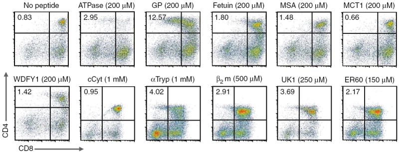
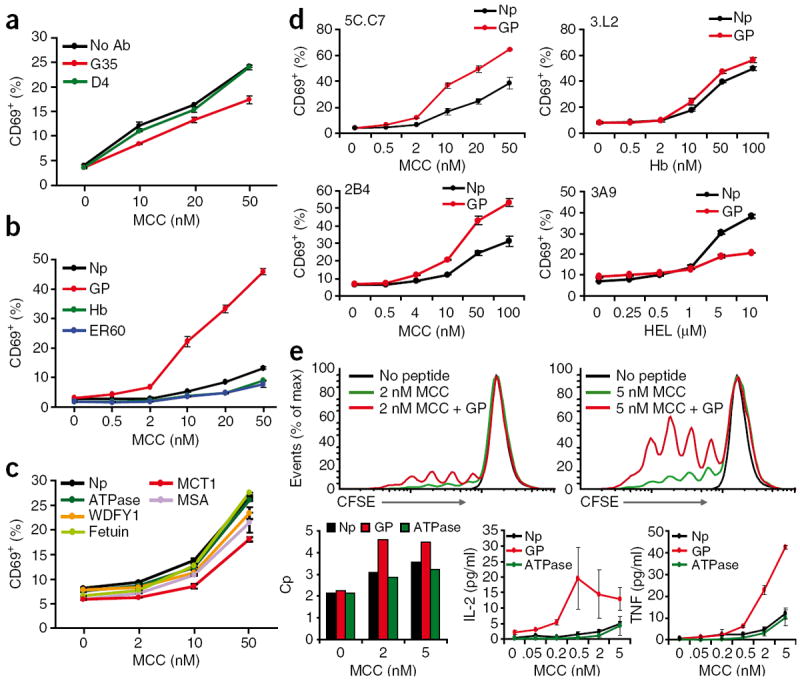
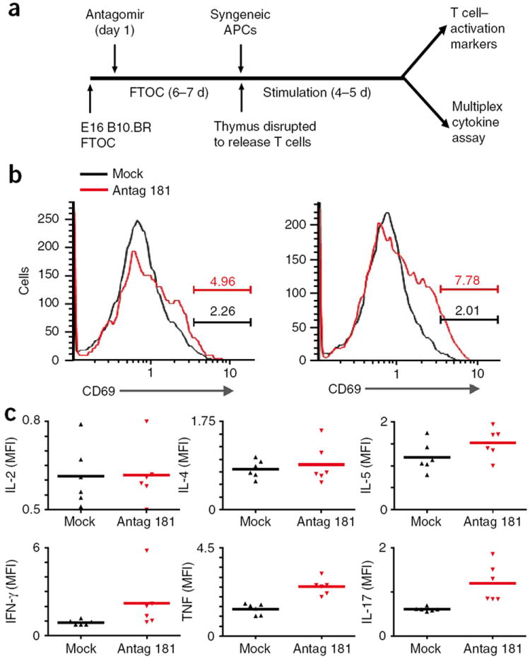
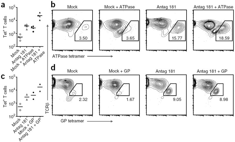
Thymic positive selection is based on the interactions of T cell antigen receptors (TCRs) with self peptide-major histocompatibility complex (MHC) ligands, but the identity of selecting peptides for MHC class II-restricted TCRs and the functional consequences of this peptide specificity are not clear. Here we identify several endogenous self peptides that positively selected the MHC class II-restricted 5C.C7 TCR. The most potent of these also enhanced mature T cell activation, which supports the hypothesis that one function of positive selection is to produce T cells that can use particular self peptide-MHC complexes for activation and/or homeostasis. We also show that inhibiting the microRNA miR-181a resulted in maturation of T cells that overtly reacted toward these erstwhile positively selecting peptides. Therefore, miR-181a helps to guarantee the clonal deletion of particular moderate-affinity clones by modulating the TCR signaling threshold of thymocytes.


Conflict of interest statement

COMPETING INTERESTS STATEMENT

The authors declare competing financial interests: details accompany the full-text HTML version of the paper at http://www.nature.com/natureimmunology/.

Figures

Figure 1
Figure 1
Positive selection of 5C.C7 T cells by endogenous peptides. Expression of CD4 and CD8 in 5C.C7 Ii-deficient FTOC cultured for 4 d with peptides in Table 1 (peptide concentrations, above plots); cultures were also analyzed for expression of CD25, CD69 and CD62L (data not shown). The cCyt, αTryp, β2m, UK1 and ER60 results are from separate experiments and are gated according to their respective no-peptide controls. MSA, mouse serum albumin; MCT1, monocarboxylate transporter 1; WDFY1, WD repeat and FYVE domain–containing 1; cCyt, complement cytolysis inhibitor; αTryp, α-anti-trypsin; β2m, β2-microglobulin; UK1, unknown 1. Data are representative of at least three independent experiments for each peptide.
Figure 2
Figure 2
An endogenous selecting peptide contributes to mature T cell activation. (a) Flow cytometry analysis of CD69 expression by naive 5C.C7 CD4+ T cells stimulated for 4 h by B10.BR splenocytes; before use as APCs, the B10.BR splenocytes were depleted of T cells, then treated with nothing (No Ab) or with antibody G35 or D4 (500 μg/ml each), washed extensively and loaded with MCC (concentration, horizontal axis). (b,c) Flow cytometry analysis of the CD69 response of naive 5C.C7 T cells stimulated for 4 h by B10.BR Ii-deficient splenocytes; before use as APCs, the B10.BR splenocytes were depleted of T cells and loaded with MCC alone (Np) or MCC plus 5 μM peptide (key; MCC concentration, horizontal axis). (d) Flow cytometry analysis of CD69 expression by 5C.C7, 3.L2, 2B4 or 3A9 TCR-transgenic T cells incubated for 5 h with B10.BR APCs plus their nominal antigenic peptides (concentration, horizontal axis) with (GP) or without (Np) 5 μM GP peptide. Hb, hemoglobin; HEL, hen egg lysozyme. (e) CFSE profiles (top row) of 5C.C7 lymph node suspensions loaded with the cytosolic dye CFSE for 10 min and washed, followed by the addition of no peptide or MCC (final concentration, 2 nM or 5 nM) with (+ GP) or without GP peptide (final concentration, 5 μM); after 4 d, cells were stained for CD4 and CD8 and assessed by flow cytometry. Bottom row, T cell–proliferative capacity (Cp; left), and supernatant concentrations of IL-2 and tumor necrosis factor (TNF) assessed by fluorescence-based cytometric bead array (bottom middle and right) of the 5C.C7 lymph node cells described above cultured with MCC (concentration, horizontal axes) plus no peptide, GP peptide or ATPase peptide. Data are representative of three (a,c,e) or five (b,d) experiments (mean and s.d.).
Figure 3
Figure 3
Importance of miR-181a for T cell central tolerance. (a) Experimental setup: E16 B10.BR FTOC were established and 1 d later were treated with antagomir 181a (35 μg/ml) or antagomir with a ‘scrambled’ seed region (mock treatment); after 6–7 total days of culture, T cells were dissociated and washed and then incubated with B10.BR (syngeneic) splenic APCs for an additional 4–5 d, then cells were stained for CD4, CD8 and CD69 and analyzed by flow cytometry (top right) and supernatants were collected for assessment of cytokine concentrations (bottom right). (b) CD69 profiles of CD4+ T cells from FTOCs treated with antagomir with a ‘scrambled’ seed region (Mock) or with miR-181a-specific antagomir (Antag 181). Numbers above bracketed lines indicate percent CD69+ cells. Data are representative of two independent experiments. (c) Fluorescence–based cytometric bead array of cytokines in supernatants from the cultures in b. Each symbol represents an individual sample; small horizontal lines indicate the mean. Data are representative of two independent experiments, each in triplicate.
Figure 4
Figure 4
Negative selection threshold set by miR-181a. Flow cytometry of cells from E16 5C.C7 TCRβ-transgenic FTOC (established as described in Fig. 3a) cultured for 7 d with antagomir 181a (35 μg/ml) or antagomir with a ‘scrambled’ seed region (Mock), then dissociated and washed and incubated for 4 d with B10.BR splenic APCs with or without 5 μM ATPase 11c peptide (a,b) or GP peptide (c,d) and then stained for CD4, CD8 and TCRβ and with ATPase–I-Ek tetramer (a,b) or GP–I-Ek tetramer (c,d). (a,c) Tetramer-positive (Tet+) T cells. Each symbol represents an independent experiment; small horizontal lines indicate the mean. (b,d) TCRβ and tetramer profiles of CD4+ T cells. Numbers in or adjacent to outlined areas indicate percent tetramer-positive cells among total CD4+ T cells. Data are representative of four experiments (ATPase 11c tetramer) or two experiments (GP tetramer).
Figure 5
Figure 5
Thymocytes downregulate miR-181a in response to selection signals. (a) Quantitative real-time PCR analysis of mature miR-181a in lysates of 5C.C7 Ii-deficient thymocytes allowed to interact for 1 h with plate-bound MCC–I-Ek or GP–I-Ek complexes or with no protein. Results are presented relative to expression in response to no protein, set as 100. Data represent two independent experiments (error bars, s.d.). (b) Quantitative real-time PCR analysis of miR-181a in DP 5C.C7 TCR–transgenic Ii-deficient thymocytes (5C.C7 Ii-KO) and 5C.C7 TCR–transgenic B10.BR thymocytes (5C.C7 B10.BR). Results are presented relative to expression in 5C.C7 Ii-KO thymocytes, set as 100. Data represent two independent experiments (error bars, s.d.).

Comment in

References

    1. Kisielow P, Bluthmann H, Staerz UD, Steinmetz M, von Boehmer H. Tolerance in T-cell-receptor transgenic mice involves deletion of nonmature CD4+8+ thymocytes. Nature. 1988;333:742–746. - PubMed
    1. Hogquist KA, Gavin MA, Bevan MJ. Positive selection of CD8+ T cells induced by major histocompatibility complex binding peptides in fetal thymic organ culture. J Exp Med. 1993;177:1469–1473. - PMC - PubMed
    1. Goldrath AW, Bevan MJ. Low-affinity ligands for the TCR drive proliferation of mature CD8+ T cells in lymphopenic hosts. Immunity. 1999;11:183–190. - PMC - PubMed
    1. Ernst B, Lee DS, Chang JM, Sprent J, Surh CD. The peptide ligands mediating positive selection in the thymus control T cell survival and homeostatic proliferation in the periphery. Immunity. 1999;11:173–181. - PubMed
    1. Stefanova I, Dorfman JR, Tsukamoto M, Germain RN. On the role of self-recognition in T cell responses to foreign antigen. Immunol Rev. 2003;191:97–106. - PubMed

Publication types