. 2009 Nov;10(11):1200-7.
doi: 10.1038/ni.1792. Epub 2009 Oct 4.

Toll-like receptor 2 on inflammatory monocytes induces type I interferon in response to viral but not bacterial ligands

Affiliations

Toll-like receptor 2 on inflammatory monocytes induces type I interferon in response to viral but not bacterial ligands

Roman Barbalat et al. Nat Immunol. 2009 Nov.

Erratum in

  • Nat Immunol. 2010 Jun;11(6):543

Abstract

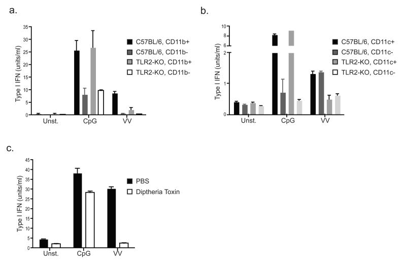
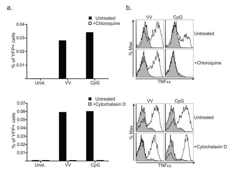
Despite the paradigm that the innate immune system uses nucleic acid-specific receptors to detect viruses because of a lack of other conserved features, many viruses are recognized by Toll-like receptor 2 (TLR2) and TLR4. The relevance of this recognition for antiviral immunity remains largely unexplained. Here we report that TLR2 activation by viruses led to the production of type I interferon. TLR2-dependent induction of type I interferon occurred only in response to viral ligands, which indicates that TLR2 is able to discriminate between pathogen classes. We demonstrate that this specialized response was mediated by Ly6C(hi) inflammatory monocytes. Thus, the innate immune system can detect certain non-nucleic acid features of viruses and links this recognition to the induction of specific antiviral genes.


Figures

Figure 1
Figure 1. TLR2-mediated recognition of vaccinia virus and MCMV
a) HEK293 cells stably transfected with a NF-κB luciferase reporter (HEK293) or the reporter together with plasmids encoding human TLR2 and human CD14 (HEK293-TLR2) were stimulated with UV-inactivated MCMV or vaccinia virus (VV) at the indicated MOIs. Pam3SK4 (Pam3) served as a positive control. Luciferase activity was measured 10 hours after activation. Fold activation was calculated relative to unstimulated cells. b & c) Bone marrow-derived DCs (b) or macrophages (c) were derived from the indicated mouse strains, stimulated with the indicated TLR ligands or viruses, and intracellular TNFα was measured by flow cytometry. Plots of stimulated cells (black lines) overlaid on plots of unstimulated cells (shaded) are shown. Plots of DCs are gated on CD11c-positive cells. The data presented are representative of at least 3 experiments.
Figure 2
Figure 2. TLR2 induces type I IFN production in response to virus
a) TLR2-dependent type I IFN production requires MyD88 but not Trif. Bone marrow cells from the indicated mice were stimulated as indicated and type I IFN was measured in the supernatants 24 hours after stimulation by bioassay. b & c) TLR2-dependent type I IFN production requires IRF3 and IRF7 but not TLR3, TLR7, or TLR9. Bone marrow cells from the indicated mice were stimulated as indicated and type I IFN production was measured by bioassay. d) TLR2 and TLR9 induce type I IFN with different kinetics. Transcripts for IFNβ and IFNα4 were measured by real-time PCR in bone marrow cells stimulated with vaccinia virus (VV) or CpG oligos. The data presented are representative of at least 2 experiments.
Figure 3
Figure 3. Differential induction of type I IFN by TLR2 in response to viral and bacterial ligands
a) IFNβ transcripts were measured in bone marrow cells treated for 12 hours with the bacterial TLR2 ligand Pam3SK4 (Pam3) or vaccinia virus (VV). b) Bone marrow or splenocytes from MOB mice were stimulated as indicated for 20 hours, and YFP production was measured by flow cytometry. The percentage of YFP-positive cells is indicated for each plot. All data are representative of at least 3 experiments.
Figure 4
Figure 4. A population of CD11b+CD11c- cells is responsible for TLR2-dependent type I IFN production
a) Bone marrow cells from C57BL/6 or TLR2-deficient mice were MACS sorted into CD11b-positive and CD11b-negative populations. The sorted cells were stimulated as indicated, and type I IFN was measured after 24 hours by bioassay. b) The same experiment was performed as described in (a), except that MACS sorting was based on CD11c. c) Splenocytes were harvested from CD11b-DTR transgenic mice 24 hours after injection with saline (PBS) or diptheria toxin. The resulting cells were stimulated as indicated and type I IFN production was measured after 24 hours by bioassay. All data are representative of at least 3 experiments.
Figure 5
Figure 5. Ly6Chigh inflammatory monocytes produce IFNβ in response to vaccinia virus
a) Bone marrow or splenocytes from MOB mice were stimulated with CpG or vaccinia virus (VV) for 20 hours, stained with antibodies against B220 or CD11c, and analyzed by flow cytometry. Comparisons between YFP-gated cells (orange line) versus total ungated cells (shaded) are shown. b) Cells were treated as in (a) but stained with antibodies against Ly6C, CD11b, or Ly6G. Comparisons between YFP-gated cells (orange line) versus total ungated cells (shaded) are shown. c) TLR2 is expressed on inflammatory monocytes. Bone marrow or splenocytes from C57BL/6 or TLR2-deficient mice were stained with antibodies specific for CD11b, Ly6C, and TLR2. d) TLR2 on inflammatory monocytes mediates type I IFN production in response to vaccinia virus. Bone marrow cells from MOB mice were cultured in the presence or absence of an anti-TLR2 blocking monoclonal antibody and stimulated as indicated. The percentage of YFP+ cells was determined by flow cytometry. e) Purified inflammatory monocytes are sufficient to produce type I IFN when stimulated with vaccinia virus. Bone marrow cells from C57BL/6 or TLR2-KO mice were sorted based on CD11b and Ly6C as shown. The plots shown have excluded B220+ and CD11c+ cells. Post-sort populations are shown and the percentage of inflammatory monocytes is indicated. The positive and negative populations were stimulated as indicated, and type I IFN production was measured after 24 hours by bioassay. All data are representative of at least 3 experiments.
Figure 6
Figure 6. Inflammatory monocytes are required for early production of type I IFN and efficient viral clearance in vivo
a) Deletion of inflammatory monocytes in CD11b-DTR mice. Splenocytes were harvested from CD11b-DTR transgenic mice 24 hours after intravenous injection of diptheria toxin or PBS followed by staining with antibodies against Ly6C, CD11b, or Ly6G. b) Inflammatory monocytes are required for production of type I IFN in response to vaccinia virus. CD11b-DTR transgenic mice where injected with diptheria toxin or PBS 24 hours before infection with 1×10⁶ PFU of vaccinia virus. Serum was collected 24 hours post infection and type I IFN were quantified by bioassay. c) Depletion of inflammatory monocytes impairs viral clearance. CD11b-DTR transgenic mice were infected with 1×10⁶ PFU of vaccinia virus 24 hours after intravenous injection of diptheria toxin or PBS. PFU were determined in the liver 48 hours after infection. The data presented are representative of at least 2 experiments.
Figure 7
Figure 7. TLR2-dependent type I IFN production requires receptor internalization
a) Bone marrow from MOB mice was incubated with 15μM chloroquine, 1μM cytochalasin D, or left untreated prior to stimulation with vaccinia virus (VV) or CpG. The percentages of YFP+ cells were measured 20h later by flow cytometry. b) Cells were treated as in (a) except that intracellular TNFα was measured in inflammatory monocytes cells by intracellular cytokine staining and flow cytometry. The data presented are representative of at least 2 experiments.

Comment in

  • TLR2 joins the interferon gang.
    Bauernfeind F, Hornung V. Bauernfeind F, et al. Nat Immunol. 2009 Nov;10(11):1139-41. doi: 10.1038/ni1109-1139. Nat Immunol. 2009. PMID: 19841644

References

    1. Medzhitov R. Recognition of microorganisms and activation of the immune response. Nature. 2007;449:819–826. - PubMed
    1. Kawai T, Akira S. Innate immune recognition of viral infection. Nature immunology. 2006;7:131–137. - PubMed
    1. Stetson DB, Medzhitov R. Type I interferons in host defense. Immunity. 2006;25:373–381. - PubMed
    1. van den Broek MF, Muller U, Huang S, Zinkernagel RM, Aguet M. Immune defence in mice lacking type I and/or type II interferon receptors. Immunological reviews. 1995;148:5–18. - PubMed
    1. Gilliet M, Cao W, Liu YJ. Plasmacytoid dendritic cells: sensing nucleic acids in viral infection and autoimmune diseases. Nature reviews. 2008;8:594–606. - PubMed

Publication types

MeSH terms