

Electrocardiographic indices of left ventricular hypertrophy and repolarization phase share the same genetic influences: a twin study
- PMID: 19804511
- PMCID: PMC6932352
- DOI: 10.1111/j.1542-474X.2009.00324.x
Electrocardiographic indices of left ventricular hypertrophy and repolarization phase share the same genetic influences: a twin study
Abstract
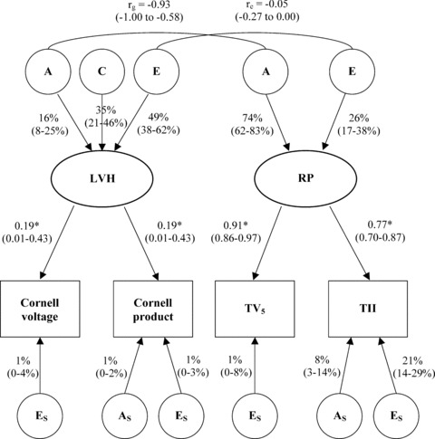
Background: Both left ventricular hypertrophy (LVH) and repolarization phase (RP) are known to be attributable to genetic influences, but less is known whether they share same genetic influences. The aim of this study was to investigate to what extent individual differences in electrocardiographic (ECG) LVH and RP are explained by genetic and environmental influences and whether these influences are shared between these two traits.
Methods: Resting ECG recordings were obtained from 186 monozygotic and 203 dizygotic female twin individuals, aged 63 to 76 years. Latent factors, called LVH and RP, were formed to condense the information obtained from LVH indices (Cornell voltage and Cornell product) and T-wave amplitudes (leads V(5) and II), respectively. Multivariate quantitative genetic modeling was used both to decompose the phenotypic variances into additive genetic, common environmental, and unique environmental influences, and for the calculation of genetic and environmental correlations between LVH and RP.
Results: Additive genetic influences explained 16% of individual differences in LVH and 74% in RP. The remaining individual differences were explained by both common and unique environmental influences. The genetic correlation and unique environmental correlation between LVH and RP were -0.93 and -0.05, respectively.
Conclusions: In older women without overt cardiac diseases, RP is under stronger genetic control than LVH. The majority of genetic influences are shared between LVH and RP whereas environmental influences are mainly specific to each.
Figures
Similar articles
-
Genetic influences on resting electrocardiographic variables in older women: a twin study.Ann Noninvasive Electrocardiol. 2009 Jan;14(1):57-64. doi: 10.1111/j.1542-474X.2008.00273.x. Ann Noninvasive Electrocardiol. 2009. PMID: 19149794 Free PMC article.
-
Diagnostic performance of electrocardiographic criteria in echocardiographic diagnosis of different patterns of left ventricular hypertrophy.Ann Noninvasive Electrocardiol. 2020 May;25(3):e12728. doi: 10.1111/anec.12728. Epub 2019 Nov 14. Ann Noninvasive Electrocardiol. 2020. PMID: 31724804 Free PMC article.
-
Baseline characteristics in relation to electrocardiographic left ventricular hypertrophy in hypertensive patients: the Losartan intervention for endpoint reduction (LIFE) in hypertension study. The Life Study Investigators.Hypertension. 2000 Nov;36(5):766-73. doi: 10.1161/01.hyp.36.5.766. Hypertension. 2000. PMID: 11082141 Clinical Trial.
-
Regression of electrocardiographic left ventricular hypertrophy or strain is associated with lower incidence of cardiovascular morbidity and mortality in hypertensive patients independent of blood pressure reduction - A LIFE review.J Electrocardiol. 2014 Sep-Oct;47(5):630-5. doi: 10.1016/j.jelectrocard.2014.07.003. Epub 2014 Jul 3. J Electrocardiol. 2014. PMID: 25052475 Review.
-
Prediction of stroke with electrocardiographic left ventricular hypertrophy in hypertensive patients: A meta-analysis.J Electrocardiol. 2020 Jul-Aug;61:27-31. doi: 10.1016/j.jelectrocard.2020.04.018. Epub 2020 Apr 28. J Electrocardiol. 2020. PMID: 32504899 Review.
Cited by
-
Heritabilities, proportions of heritabilities explained by GWAS findings, and implications of cross-phenotype effects on PR interval.Hum Genet. 2015 Nov;134(11-12):1211-9. doi: 10.1007/s00439-015-1595-9. Epub 2015 Sep 18. Hum Genet. 2015. PMID: 26385552 Free PMC article.
References
-
- Levy D, Anderson KM, Savage DD, et al Echocardiographically detected left ventricular hypertrophy: Prevalence and risk factors. The Framingham Heart Study. Ann Intern Med 1988;108:7–13. - PubMed
-
- Liebson PR, Grandits G, Prineas R, et al Echocardiographic correlates of left ventricular structure among 844 mildly hypertensive men and women in the Treatment of Mild Hypertension Study (TOMHS). Circulation 1993;87:476–486. - PubMed
-
- Vakili BA, Okin PM, Devereux RB. Prognostic implications of left ventricular hypertrophy. Am Heart J 2001;141:334–341. - PubMed
-
- Carter WA, Estes EH Jr. Electrocardiographic manifestations of ventricular hypertrophy: A computer study of ECG‐anatomic correlations in 319 cases. Am Heart J 1964;68:173–182. - PubMed
-
- Devereux RB, Reichek N. Repolarization abnormalities of left ventricular hypertrophy. Clinical, echocardiographic and hemodynamic correlates. J Electrocardiol 1982;15:47–53. - PubMed
Publication types
MeSH terms
LinkOut - more resources
Full Text Sources
