. 2009;11(5):R149.
doi: 10.1186/ar2823. Epub 2009 Oct 5.

Chemokine receptor expression and functional effects of chemokines on B cells: implication in the pathogenesis of rheumatoid arthritis

Affiliations

Chemokine receptor expression and functional effects of chemokines on B cells: implication in the pathogenesis of rheumatoid arthritis

Toshihiro Nanki et al. Arthritis Res Ther. 2009.

Abstract

Introduction: Accumulation of B cells in the rheumatoid arthritis (RA) synovium has been reported, and it has been thought that these cells might contribute to the pathogenesis of RA by antigen presentation, autoantibody production, and/or inflammatory cytokine production. Chemokines could enhance the accumulation of B cells in the synovium. The aims of this study were to determine chemokine receptor expression by B cells both in the peripheral blood of normal donors and subjects with RA, and at the inflammatory site in RA, and the effects of chemokines on B cell activation.

Methods: Cell surface molecule expression was analyzed by flow cytometry. Cellular migration was assessed using chemotaxis chambers. Cellular proliferation was examined by 3H-thymidine incorporation. Tumor necrosis factor (TNF) production was assayed by enzyme-linked immunosorbent assay.

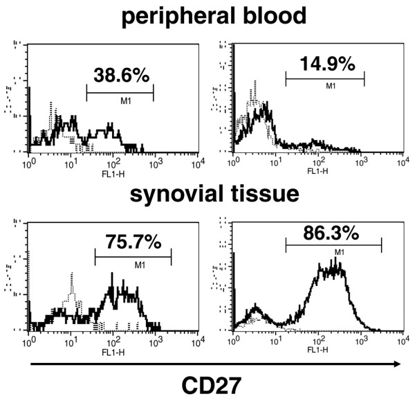
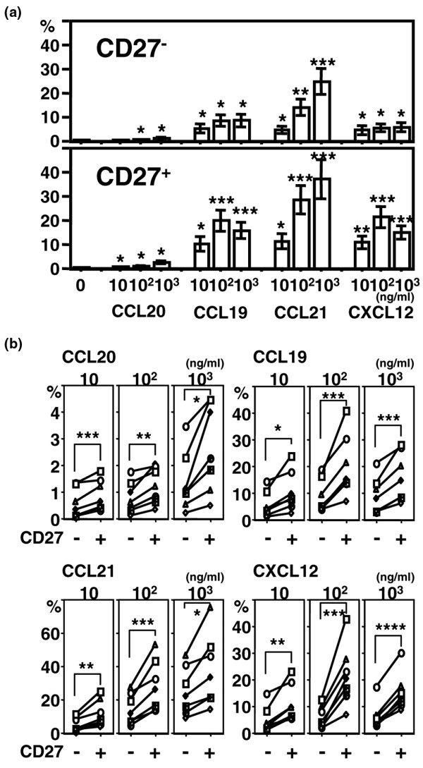
Results: Significant numbers of peripheral blood B cells of healthy donors and subjects with RA expressed CC chemokine receptor (CCR)5 and CXCR3, and most B cells expressed CCR6, CCR7, CXCR4 and CXCR5. CCR5 expression was more frequent on CD27+ than CD27- peripheral blood B cells of healthy donors and RA. Synovial B cells more frequently expressed CCR5, but less often expressed CCR6, CCR7 and CXCR5 compared to peripheral blood in RA. Further functional analyses were performed on peripheral blood B cells from healthy donors. Migration of peripheral blood B cells, especially CD27+ B cells, was enhanced by CC chemokine ligand (CCL)20, CCL19, CCL21 and CXCL12. All four chemokines alone induced B cell proliferation; with CCL21 being the most effective. CCL21 also enhanced the proliferation of anti-immunoglobulin (Ig)M-stimulated B cells and blockade of CCR7 inhibited this effect. CCL20, CCL21 and CXCL12 enhanced TNF production by anti-IgM mAb-stimulated B cells. Finally, stimulation with CXCL12, but not CCL20, CCL19 and CCL21, enhanced inducible costimulator-ligand (ICOSL) expression by peripheral blood B cells of healthy donors and RA, but did not increase B cell-activating factor receptor or transmembrane activator and CAML-interactor.

Conclusions: The data suggest that CCR5, CCR6, CCR7, CXCR3, CXCR4 and CXCR5 may be important for the B cell migration into the synovium of RA patients, and also their local proliferation, cytokine production and ICOSL expression in the synovium.


Figures

Figure 1
Figure 1
Chemokine receptor expression by B cells. Peripheral blood mononuclear cells (PBMCs) from healthy donors (n = 4 to 7) and rheumatoid arthritis (RA) patients (n = 17 to 18) and synovial cells from RA patients (n = 10 to 11) were stained with CD19, CD27, and CCR5, CCR6, CCR7, CXCR3, CXCR4 or CXCR5, and the expression of the various markers was analyzed by flow cytometry. CD19+ B cells were gated, and the frequency of expression of each chemokine receptor is shown. Data represent mean ± standard error of the mean. *P < 0.05, **P < 0.01, ***P < 0.001, ****P < 0.0001. ST = synovial tissue.
Figure 2
Figure 2
CD27 expression by peripheral blood and synovial tissue B cells of subjects with RA. CD19+ B cells were gated, and representative histograms from two patients with rheumatoid arthritis (RA) show the cells stained with anti-CD27 monoclonal antibody (mAb) (solid lines) and isotype-matched control (dotted lines).
Figure 3
Figure 3
B cell migration in response to chemokines. Peripheral blood mononuclear cells (PMBCs) from healthy donors were cultured in the presence of various concentrations of CCL20, CCL19, CCL21, or CXCL12 for two hours. The cells migrated through ECV304-coated transwells were stained with CD19 and CD27, and the numbers of cells were assessed. The percentage of migrated cells was calculated by dividing the number of migrated CD27- or CD27+ B cells by the number of total cultured CD27- or CD27+ B cells for six to seven donors. (a) Values are mean ± standard error of the mean. *P < 0.05, **P < 0.01, ***P < 0.005, vs no chemokine. (b) Each symbol represents an individual subject. *P < 0.05, **P < 0.01, ***P < 0.005, ****P < 0.0001.
Figure 4
Figure 4
B cell proliferation in response to chemokine stimulation. Purified B cells from peripheral blood mononuclear cells (PBMCs) of normal donors were stimulated with the indicated chemokines for 48 hours (a) without and (b) with anti-IgM stimulation. (c) To block CCR7, the B cells were pre-incubated with anti-CCR7 monoclonal antibody (mAb) or control mAb for 30 minutes. 3H-thymidine was added and B cells were incubated for 24 hours. The incorporated radioactivity was quantified. Fold increase in 3H-thymidine incorporation in response to chemokine stimulation for four to eight donors was calculated. Values are mean ± standard error of the mean. (a, b) *P < 0.05, **P < 0.005, ***P < 0.0005, vs no chemokine stimulation. (c) *P < 0.05, **P < 0.005.
Figure 5
Figure 5
TNF production by chemokine stimulation. Purified peripheral blood B cells from normal donors were incubated with the indicated chemokines with or without anti-IgM monoclonal antibody (mAb) for 24 hours. The concentration of TNF in the culture supernatant was measured by ELISA. Data are mean ± standard error of the mean values of three independent experiments analyzed in duplicate. *P < 0.05, **P < 0.005, ***P < 0.0005, vs no chemokine stimulation.
Figure 6
Figure 6
Cell surface expression of ICOSL and BAFF receptors. Peripheral blood mononuclear cells (PBMCs) from (a) healthy donors and (b) subjects with rheumatoid arthritis (RA) were stimulated with the indicated chemokines for 24 hours. Afterward, the cells were stained with monoclonal antibody (mAbs) to CD19 and inducible costimulator-ligand (ICOSL), B cell-activating factor receptor (BAFF-R) or transmembrane activator and CAML-interactor (TACI), and the expression was analyzed by flow cytometry. Representative expression patterns by CD19+ cells are shown from three similar independent experiments.

References

    1. Edwards JC, Cambridge G. Sustained improvement in rheumatoid arthritis following a protocol designed to deplete B lymphocytes. Rheumatology (Oxford) 2001;40:205–211. doi: 10.1093/rheumatology/40.2.205. - DOI - PubMed
    1. Cohen SB, Emery P, Greenwald MW, Dougados M, Furie RA, Genovese MC, Keystone EC, Loveless JE, Burmester GR, Cravets MW, Hessey EW, Shaw T, Totoritis MC. Rituximab for rheumatoid arthritis refractory to anti-tumor necrosis factor therapy: Results of a multicenter, randomized, double-blind, placebo-controlled, phase III trial evaluating primary efficacy and safety at twenty-four weeks. Arthritis Rheum. 2006;54:2793–2806. doi: 10.1002/art.22025. - DOI - PubMed
    1. Lundy SK, Sarkar S, Tesmer LA, Fox DA. Cells of the synovium in rheumatoid arthritis. T lymphocytes. Arthritis Res Ther. 2007;9:202. doi: 10.1186/ar2107. - DOI - PMC - PubMed
    1. Kinne RW, Stuhlmuller B, Burmester GR. Cells of the synovium in rheumatoid arthritis. Macrophages. Arthritis Res Ther. 2007;9:224. doi: 10.1186/ar2333. - DOI - PMC - PubMed
    1. Lutzky V, Hannawi S, Thomas R. Cells of the synovium in rheumatoid arthritis. Dendritic cells. Arthritis Res Ther. 2007;9:219. doi: 10.1186/ar2200. - DOI - PMC - PubMed

Publication types

MeSH terms

Substances