. 2009 Oct 5:2:31.
doi: 10.1186/1756-6606-2-31.

NADPH oxidase mediates beta-amyloid peptide-induced activation of ERK in hippocampal organotypic cultures

Affiliations

NADPH oxidase mediates beta-amyloid peptide-induced activation of ERK in hippocampal organotypic cultures

Faridis Serrano et al. Mol Brain. .

Abstract

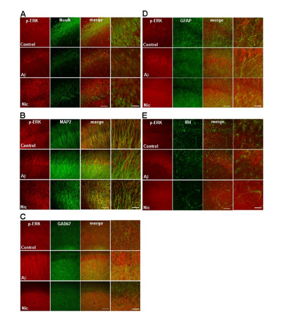
Background: Previous studies have shown that beta amyloid (Abeta) peptide triggers the activation of several signal transduction cascades in the hippocampus, including the extracellular signal-regulated kinase (ERK) cascade. In this study we sought to characterize the cellular localization of phosphorylated, active ERK in organotypic hippocampal cultures after acute exposure to either Abeta (1-42) or nicotine.

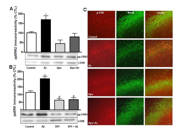
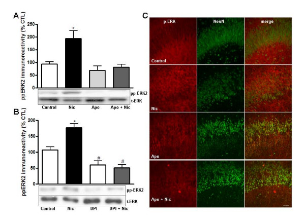
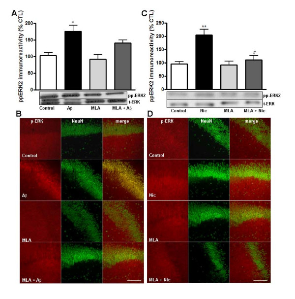
Results: We observed that Abeta and nicotine increased the levels of active ERK in distinct cellular localizations. We also examined whether phospho-ERK was regulated by redox signaling mechanisms and found that increases in active ERK induced by Abeta and nicotine were blocked by inhibitors of NADPH oxidase.

Conclusion: Our findings indicate that NADPH oxidase-dependent redox signaling is required for Abeta-induced activation of ERK, and suggest a similar mechanism may occur during early stages of Alzheimer's disease.


Figures

Figure 1
Figure 1
Aβ- and nicotine-induced activation of ERK in cultured hippocampal slices. Hippocampal slice cultures were treated with culture media, media containing Aβ (100 nM), or media containing nicotine (500 nM) for either 5 min or 10 min, respectively. Confocal images of hippocampal area CA1 were obtained from slices that were double labeled using antibodies specific for phospho-ERK (red) and A) the neuronal marker NeuN (green), B) the dendritic marker MAP2 (green), C) a marker for GABAergic interneurons GAD67 (green), D) the astrocytic marker GFAP (green), and E) the microglial marker IB4 (green). Dual labeling is indicated by yellow/orange. All the images were taken with a 20× objective and inserts represent higher magnifications (optical zoom 3.0). The scale bars are the same in all images and represent 20 μm and 10 μm, respectively.
Figure 2
Figure 2
Aβ peptide-induced activation of ERK is blocked by the NADPH oxidase inhibitors apocynin and DPI. Hippocampal slice cultures were pretreated for either 25 or 55 min with media in the presence or absence of either 100 μM apocynin (A) or 10 μM DPI (B), followed by incubation with Aβ (100 nM, 5 min). A, B) Western blots. Phospho-ERK immunoreactivity was normalized to total ERK and expressed as percent of control. Data represent the mean ± SEM; n = 7-10; *p < 0.01; **p < 0.001; #p < 0.05. C) Immunocytochemistry. Confocal images of hippocampal area CA1 were obtained from slices that were double labeled using antibodies specific for phospho-ERK (red) and the neuronal marker NeuN (green). Dual labeling is indicated by yellow/orange. Images were taken with a 20× objective and the scale bar represents 20 μm.
Figure 3
Figure 3
Nicotine-induced activation of ERK is blocked by the NADPH oxidase inhibitors apocynin and DPI. Hippocampal slice cultures were pretreated for 25 or 55 min with media in the presence or absence of either 100 μM apocynin (A) or 10 μM DPI (B), followed by incubation with nicotine (500 nM, 10 min). A, B) Western blots. Phospho-ERK immunoreactivity was normalized to total ERK and expressed as percent of control. Data represent the mean ± SEM; n = 6-8; * p < 0.01, ** p < 0.001. C) Immunocytochemistry. Confocal images of hippocampal area CA1 were obtained from slices that were double labeled using antibodies specific for phospho-ERK (red) and the neuronal marker NeuN (green). Dual labeling is indicated by yellow/orange. Images were taken with a 20× objective and the scale bar represents 20 μm.
Figure 4
Figure 4
Aβ- and nicotine-induced activation of ERK is decreased by the α7nAChR inhibitor MLA. Hippocampal slice cultures were pretreated for 30 min with media in the presence or absence of MLA (10 nM) followed by incubation with Aβ (100 nM, 5 min). A) Western blots. Phospho-ERK immunoreactivity was normalized to total ERK and expressed as percent of control. Data represent the mean ± SEM. n = 3-6; *p < 0.01. B) Immunocytochemistry. Confocal images were obtained from slices that were double labeled using antibodies specific for phospho-ERK (red) and the neuronal marker NeuN (green). Dual labeling is indicated by yellow/orange. Hippocampal slice cultures were pretreated for 30 min with media in the presence or absence of MLA (10 nM) followed by incubation with nicotine (500 nM, 5 min). C) Western blots. Phospho-ERK immunoreactivity was normalized to total ERK and expressed as percent of control. Data represent the mean ± SEM. n = 3-6, **p < 0.001. D) Immunocytochemistry. Confocal images were obtained from slices that were double labeled using antibodies specific for phospho-ERK (red) and the neuronal marker NeuN (green). Dual labeling is indicated by yellow/orange. Images were taken with a 20× objective and the scale bar represents 50 μm.

References

    1. Kuo YM, Emmerling MR, Vigo-Pelfrey C, Kasunic TC, Kirkpatrick JB, Murdoch GH, Ball MJ, Roher AE. Water-soluble Abeta (N-40, N-42) oligomers in normal and Alzheimer disease brains. J Biol Chem. 1996;271:4077–4081. doi: 10.1074/jbc.271.8.4077. - DOI - PubMed
    1. Kamenetz F, Tomita T, Hsieh H, Seabrook G, Borchelt D, Iwatsubo T, Sisodia S, Malinow R. APP processing and synaptic function. Neuron. 2003;37:925–937. doi: 10.1016/S0896-6273(03)00124-7. - DOI - PubMed
    1. Bell KA, O'Riordan KJ, Sweatt JD, Dineley KT. MAPK recruitment by beta-amyloid in organotypic hippocampal slice cultures depends on physical state and exposure time. J Neurochem. 2004;91:349–361. doi: 10.1111/j.1471-4159.2004.02722.x. - DOI - PubMed
    1. Dineley KT, Westerman M, Bui D, Bell K, Ashe KH, Sweatt JD. Beta-amyloid activates the mitogen-activated protein kinase cascade via hippocampal alpha7 nicotinic acetylcholine receptors: In vitro and in vivo mechanisms related to Alzheimer's disease. J Neurosci. 2001;21:4125–4133. - PMC - PubMed
    1. Dineley KT, Bell KA, Bui D, Sweatt JD. beta-Amyloid peptide activates alpha 7 nicotinic acetylcholine receptors expressed in Xenopus oocytes. J Biol Chem. 2002;277:25056–25061. doi: 10.1074/jbc.M200066200. - DOI - PubMed

Publication types

MeSH terms

LinkOut - more resources