. 2009 Oct 7;97(7):1997-2003.
doi: 10.1016/j.bpj.2009.06.051.

Protein-mediated molecular bridging: a key mechanism in biopolymer organization

Affiliations

Protein-mediated molecular bridging: a key mechanism in biopolymer organization

Paul A Wiggins et al. Biophys J. .

Abstract

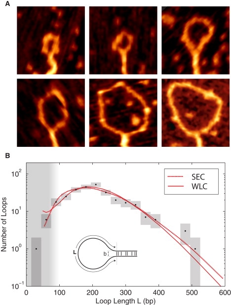
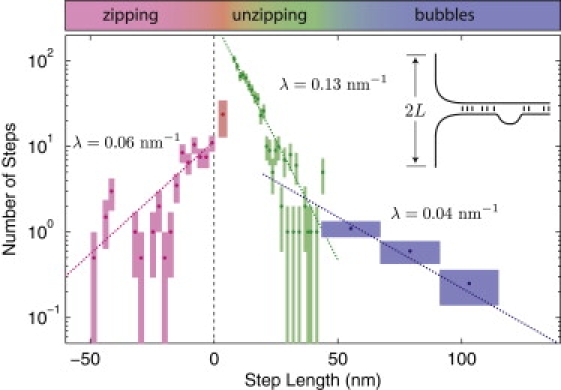
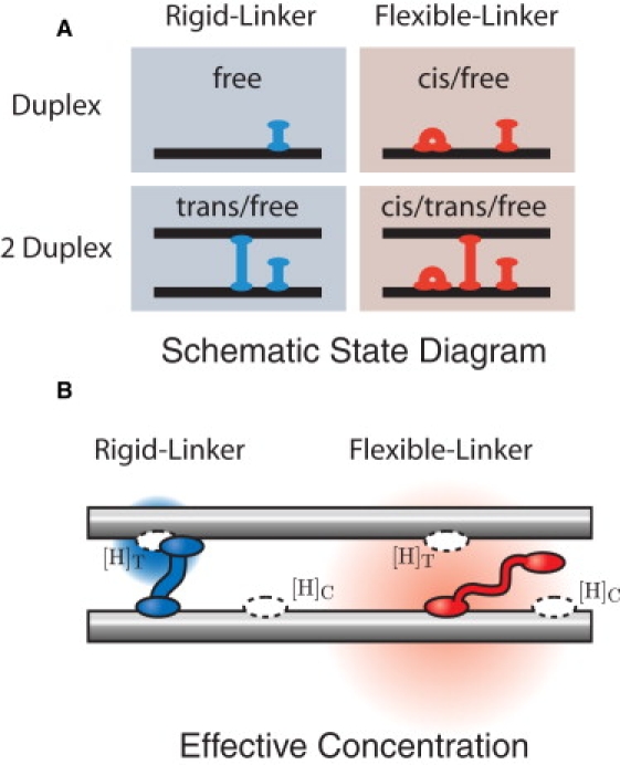
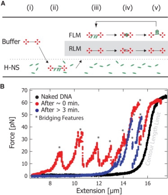
Protein-mediated bridging is ubiquitous and essential for shaping cellular structures in all organisms. Here we dissect this mechanism for a model system: the Histone-like Nucleoid-Structuring protein (H-NS). We present data from two complementary single-molecule assays that probe the H-NS-DNA interaction: a dynamic optical-trap-driven unzipping assay and an equilibrium H-NS-mediated DNA looping scanning force microscopy imaging assay. To quantitatively analyze and compare these assays, we employ what we consider a novel theoretical framework that describes the bridging motif. The interplay between the experiments and our theoretical model not only infers the effective interaction free energy, the bridging conformation and the duplex-duplex spacing, but also reveals a second, unresolved, cis-binding mode that challenges our current understanding of the role of bridging proteins in chromatin structure. We expect that this theoretical framework for describing protein-mediated bridging will be applicable to proteins acting in chromatin and cytoskeletal organization.


Figures

Figure 1
Figure 1
(A) Representative SFM images of H-NS-induced DNA loop complexes. The images are 125 nm by 125 nm. (B) The probability of H-NS-mediated DNA loops length L. A fit of the observed loop-length distribution (black), determined from SFM images of 286 loops, to the predicted loop-length distribution (red) results in excellent agreement with both chain statistics models. These fits predict nearly identical values for the adhesion energy: ρ = 0.13 ± 0.03 kT nm−1 (SEC) and ρ = 0.14 ± 0.03 kT nm−1 (WLC). The SEC model predicts a loop spacing of b = 4 ± 1 nm, whereas the WLC suggests a slightly higher value of b = 6 ± 1 nm . The chain statistical models are discussed in the Supporting Material. (The vertical range of the boxes represents the Poisson error, and the width represents the bin size.)
Figure 2
Figure 2
Step-size distribution for saturated, bridged duplexes upon optical-tweezers driven unzipping. In this assay, the step lengths are determined by the bridge spacing in the DNA–H-NS–DNA complex. The number of observed contour length steps is plotted as a function of the step length. The Poisson error is represented by the vertical box range. The bin size is represented by the box width. (Where the bin size has been expanded, the observed event number has been renormalized, resulting in fractional events.) The first step bridging probability (red) appears suppressed relative to the amplitude of subsequent steps in the unzipping regime which are well described by an exponential decay (green). The decay length fit from n > 1 steps is λ = 0.13 ± 0.02 nm−1.
Figure 3
Figure 3
(A) Schematic state diagram of H-NS–DNA interactions. In our schematic drawings, we represent the H-NS dimer as a single unit. We consider two simple models for H-NS–DNA interaction: the Rigid and Flexible-Linker models. In the Flexible-Linker model, the H-NS dimer can bind to DNA in three states: free-head, trans, and cis. When H-NS interacts with two DNA duplexes, there is a competition between the three modes of binding. DNA adhesion is driven only by entropic effects (since H-NS can assume the cis conformation at the same energetic cost). In the Rigid-Linker model, the linker is too stiff to efficiently permit cis binding and the H-NS–DNA interaction is dominated by the free-head and trans binding modes. (B) The effective concentration illustrated for the Flexible and Rigid-Linker models. When H-NS binds a head to a DNA duplex, the diffusion of the second head is constrained. The physical concentration ([H]) of the second head in the proximal volume is dramatically increased. (The concentration is schematically illustrated by the red and blue gradients where the deeper hue corresponds to a higher local concentration.) This effective concentration is predicted by a polymer model for the linker domain. Structural and mutational studies of the H-NS protein suggest that the dimerization and DNA-binding domains are connected by a flexible linker (15). The Flexible-Linker model makes use of the fact that the linker domain can be modeled as a Gaussian chain with the generic amino-acid Kuhn length. In contrast, the linker domain of the Rigid-Linker model is assumed to have a structure which localizes the head domain in proximity to the trans-binding site. We therefore expect the trans head concentration to be much greater than that computed in the Flexible-Linker model.
Figure 4
Figure 4
H-NS-DNA complex lifetime experiment. (A) A single DNA molecule is caught with optical tweezers. A force extension curve is taken in buffer solution (i). Subsequently, the DNA is “loaded” with H-NS in a buffer containing H-NS (2 μM) (ii). The DNA molecule is then placed back into the buffer solution without H-NS (iii). To probe the occurrence of bridge formation (and thus detect the presence of H-NS), the beads were brought together, releasing the DNA tension and allowing trans binding (iv). The force-extension curves were taken at time intervals of 1–5 min (ivviii). The Rigid-Linker model (gray box) predicts that when the DNA is extended (iii), the lifetime of the H-NS-DNA complex is the lifetime of a single head (∼1 s). The Flexible-Linker model predicts that when the DNA molecule is extended (iii), the lifetime of the H-NS-DNA complex will be on order minutes due to the cooperative binding of the two heads. (B) The black curve shows the initial force-extension response of naked DNA. The red curve shows the force-extension response for the DNA immediately after incubation with H-NS. Bridging features are force spikes () at extensions shorter than the contour length that result from large regions of trans-bound H-NS. After holding an extended state for >3 min, the blue curve still exhibits bridging features () that demonstrate that H-NS remains bound to DNA for a lifetime an order of magnitude greater than predicted by the single-head off rate. These data strongly support the existence of cis-bound H-NS as predicted by the Flexible-Linker model.

References

    1. Dorman C.J. H-NS: a universal regulator for a dynamic genome. Nat. Rev. Microbiol. 2004;2:391–400. - PubMed
    1. Dorman C.J. H-NS, the genome sentinel. Nat. Rev. Microbiol. 2007;5:157–161. - PubMed
    1. Dame R.T., Noom M.C., Wuite G.J.L. Bacterial chromatin organization by H-NS protein unravelled using dual DNA manipulation. Nature. 2006;444:387–390. - PubMed
    1. Dame R.T. The role of nucleoid-associated proteins in the organization and compaction of bacterial chromatin. Mol. Microbiol. 2005;56:858–870. - PubMed
    1. Luijsterburg M.S., Noom M.C., Wuite G.J.L., Dame R.T. The architectural role of nucleoid-associated proteins in the organization of bacterial chromatin: a molecular perspective. J. Struct. Biol. 2006;156:262–272. - PubMed

Publication types

LinkOut - more resources