. 2009 Oct 2;139(1):161-74.
doi: 10.1016/j.cell.2009.08.027.

Stringent specificity in the construction of a GABAergic presynaptic inhibitory circuit

Affiliations

Stringent specificity in the construction of a GABAergic presynaptic inhibitory circuit

J Nicholas Betley et al. Cell. .

Abstract

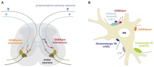
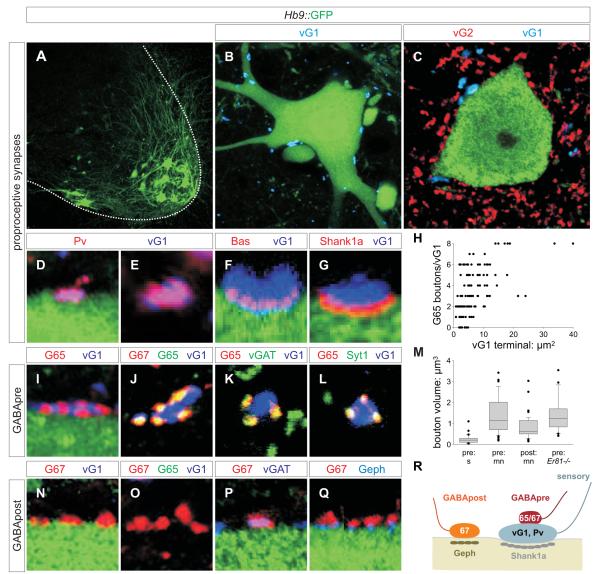
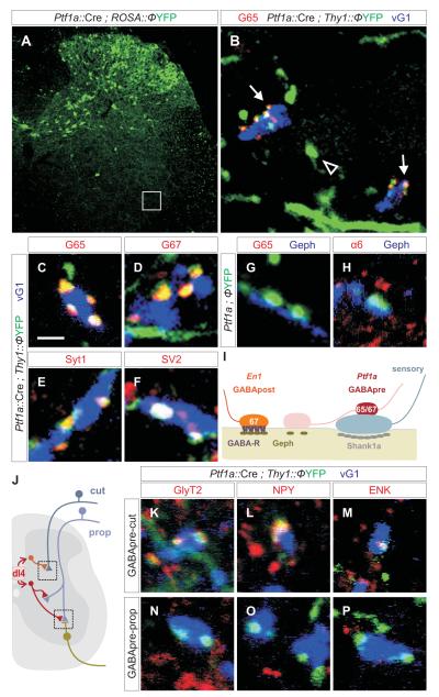
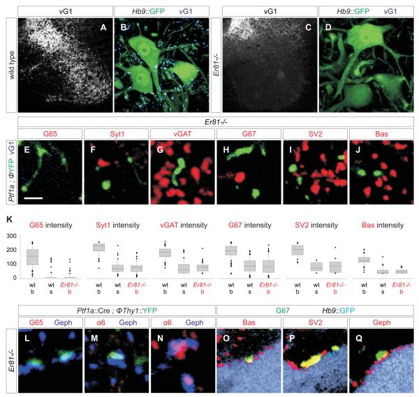
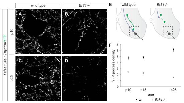
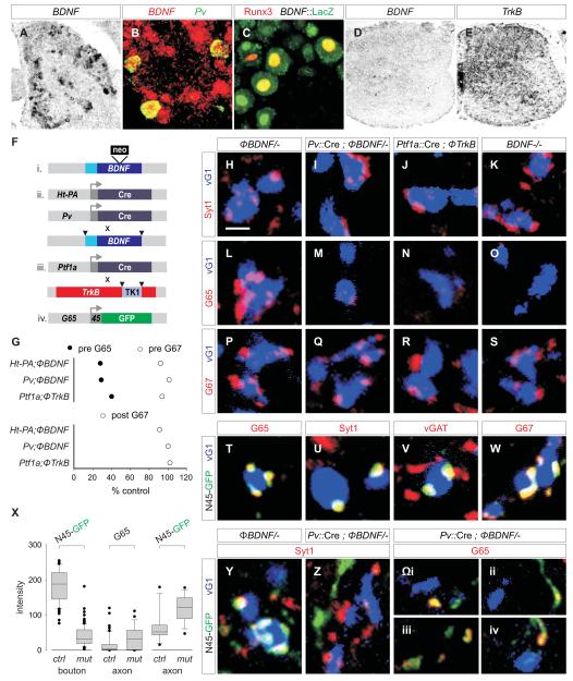
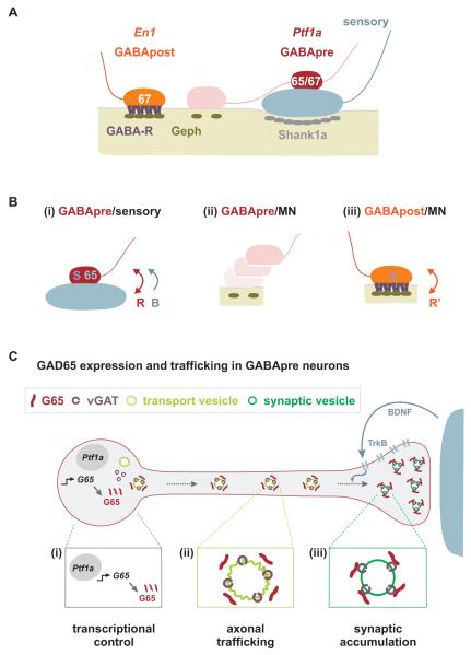
GABAergic interneurons are key elements in neural coding, but the mechanisms that assemble inhibitory circuits remain unclear. In the spinal cord, the transfer of sensory signals to motor neurons is filtered by GABAergic interneurons that act presynaptically to inhibit sensory transmitter release and postsynaptically to inhibit motor neuron excitability. We show here that the connectivity and synaptic differentiation of GABAergic interneurons that mediate presynaptic inhibition is directed by their sensory targets. In the absence of sensory terminals these GABAergic neurons shun other available targets, fail to undergo presynaptic differentiation, and withdraw axons from the ventral spinal cord. A sensory-specific source of brain derived neurotrophic factor induces synaptic expression of the GABA synthetic enzyme GAD65--a defining biochemical feature of this set of interneurons. The organization of a GABAergic circuit that mediates presynaptic inhibition in the mammalian CNS is therefore controlled by a stringent program of sensory recognition and signaling.


Figures

Figure 1
Figure 1. Two GABAergic inhibitory circuits in the spinal cord
(A) Circuitry of GABApre and GABApost neurons. (B) Synaptic inputs on motor neurons include vGlut1 (vG1)+ proprioceptive sensory terminals, GABApost terminals, vGlut2 (vG2)+ interneuron terminals, vAChT+ cholinergic interneuron terminals, and corticospinal terminals (CST). Propioceptive terminals are contacted by GABApre terminals.
Figure 2
Figure 2. Inhibitory microcircuitry at sensory-motor synapses
(A) GFP+ motor neurons. (B, C) GFP+ motor neurons contacted by vGlut1 (vG1)+ and vGlut2 (vG2)+ terminals. (D, E) vG1+ terminals on motor neurons co-express Pv at p7. 95.6 ± 0.9% vG1+ terminals are labeled with mGFP in a Pv::Cre ; Tau::ΦmGFP cross, indicating their sensory origin (Figure S1A-C) (for all quantitations herein, data are represented as mean +/− s.e.m.; n = 971 vG1+ boutons; 3 mice). (F) vG1+ terminals express Bas at sites of contact with GFP+ motor neurons. (G) 78.8 ± 2.0% of vG1+ terminals (n = 259 boutons; 2 mice) on motor neurons align with post-synaptic Shank1a. (H) GAD65 (G65)+ boutons / vG1+ terminal as a function of surface area of the vG1+ terminal, at p15 to 21. 89.9 ± 3.1% (n = 381 boutons; 2 mice) of vG1+ terminals were contacted by at least one G65+ bouton (n = 126 vG1+ terminals, 3 mice). (I-L) GAD expression by GABApre boutons. 99.8 ± 0.2% (n = 746 boutons; 4 mice) of G65+ terminals contact vG1+ sensory terminals. >99% of G65+ boutons express GAD67 (G67), vGAT and Syt1. (M) Distribution of the volumes of GABAergic boutons and varicosities in ventral spinal cord. Values obtained from >100 bouton measurements in ≥ 3 mice. In each comparison, except between pre: mn contacts in the wild type and Er81-/- background, differences are significant at p <0.0001 (Mann-Whitney U Test). (N-Q) Synaptic marker expression at GABApost synapses. G67+, G65off terminals contact GFP+ motor neurons, independent of proximity to vG1+ sensory terminals. G67+ terminals express vGAT. 96.8 ± 1.8% (n = 339 boutons; 3 mice) of G67+, G65off terminals are aligned with motor neuron Geph+ puncta. (R) Inhibitory synaptic organization at sensory-motor synapses. Images from rostral lumbar levels of p21 Hb9::GFP mice unless indicated.
Figure 3
Figure 3. Distinct classes of Ptf1a-derived GABApre neurons target proprioceptive and cutaneous sensory afferent terminals
(A) Dorsal restriction of YFP+ neurons in Ptf1a::Cre; Thy1::ΦYFP mice. (B) Ptf1a::Cre ; Thy1::ΦYFP marked axons and terminals (subset of box in A). Thy1::YFP+ axons (arrowheads) are observed in the vicinity of motor neurons. GAD65 (G65) expression is restricted to compact axonal boutons (arrows) on vGlut1 (vG1)+ terminals. (C-F) Ptf1a::Cre ; Thy1::ΦYFP-labeled boutons on vG1+ terminals express G65 (C), G67 (D), Syt1 (E) and SV2 (F). In Ptf1a::Cre ; Tau::ΦmGFP mice, 90.9 ± 6.2% (n = 465 boutons; 3 mice) of G65+ synapses on vG1+ sensory terminals express mGFP. In many domains of the ventral horn ~100% of GABApre terminals express mGFP, suggesting G65+, mGFPoff terminals result from inefficient Cre recombination. (G, H) About 50% of Ptf1a::Cre ; Thy1::ΦYFP-labeled GABApre axonal varicosities (see Figure 2M for volume) align with post-synaptic Geph+ puncta but do not align with GABAA receptor α6 or β2/3 subunits (H, Figure S9). These motor neuron-associated YFP+ varicosities do not express G65 (G), G67, Syt1, Bas or SV2 (Figure S7A; data not shown). (I) Neuronal target directs the differentiation of Ptf1a-marked GABApre boutons. (J) Ptf1a-derived Di4 interneurons form synapses with cutaneous afferent terminals in the dorsal spinal cord and proprioceptive terminals in the ventral horn. (K-M) In p15 Ptf1a::Cre ; Thy1::ΦYFP mice, GABApre terminals synapse with vGlut1 (vG1)+ cutaneous sensory terminals and express GlyT2 (K), NPY (L), and ENK (M). (N-P) In p15 Ptf1a::Cre ; Thy1::ΦYFP mice, GABApre terminals synapse with vG1+ proprioceptive terminals, but do not express GlyT2 (N), NPY (O), or ENK (P). Images from lumbar spinal cord of p21 mice. Scale bar: 2 μm in C-H, K-P.
Figure 4
Figure 4. GABApre synaptic differentiation fails in the absence of sensory terminals
(A-D) vGlut1 expression in wild type (A, B) and Er81-/- (C, D) spinal cord. (E-J) Synaptic marker expression in YFP+ GABApre axons and varicosities in the ventral horn of Ptf1a::Cre ; Thy1::ΦYFP ; Er81-/- mice. (K) Synaptic protein levels in YFP+ axon shafts (s) and boutons (b) in Ptf1a::Cre ; Thy1::ΦYFP (wt) and Ptf1a::Cre ; Thy1::ΦYFP ; Er81-/- (Er81-/-) mice. Differences in protein level between wild type and Er81-/- GABApre boutons/varicosities are significant at p <0.0001 (Mann-Whitney U Test). (L-N) In Ptf1a::Cre ; Thy1::ΦYFP ; Er81-/- mice, YFP+ contacts on motor neurons are aligned with Geph+ puncta, but are large and lack pre-synaptic G65 (L) and post-synaptic GABAA α6 (M). Geph+ puncta lacking pre-synaptic YFP (GABApost) accumulate post-synaptic GABAA α6 (N). (O-Q) In Hb9::GFP ; Er81-/- mice, G67+ terminals (green) contact GFP+ motor neurons (light blue), co-express Bas (O), SV2 (P) and vGAT (not shown), and align with motor neuron Geph+ puncta (Q). Images from p15 mice. Scale bar: 2 μm in E-J, L-Q.
Figure 5
Figure 5. Retraction of GABApre axons in the absence of sensory terminals
(A-D) YFP+ axons in the ventral horn of Ptf1a::Cre ; Thy1::ΦYFP (A, C) and Ptf1a::Cre ; Thy1::ΦYFP ; Er81-/- (B, D) mice. Images are 3 μm z-stacks from region marked in E. (E) Retraction of GABApre axons in Er81-/- mice (motor neurons in grey, sensory afferents in blue and GABApre axons in green). (F) Density of YFP+ axons and boutons in the ventral horn of Ptf1a::Cre ; Thy1::ΦYFP (wt) and Ptf1a::Cre ; Thy1::ΦYFP ; Er81-/- (Er81-/-) mice at p10, p15 and p25. See Supplementary Procedures for quantitation.
Figure 6
Figure 6. Accumulation of GAD65 in GABApre boutons depends on sensory-derived BDNF signaling
(A) 30.5 ± 0.7% (n = 464 neurons; 3 mice) of p7 lumbar DRG neurons express BDNF mRNA. (B) ~85% (n = 3 mice) of p7 Pv+ proprioceptive neurons express BDNF mRNA. (C) 81.5 ± 3.8% (n = 54 neurons; 2 mice) of Runx3+ proprioceptive DRG neurons express LacZ in BDNF::LacZ reporter mice. (D) BDNF expression in p7 spinal cord. (E) TrkB expression in p7 spinal cord. (F) Cre driver lines, BDNF, and TrkB alleles used. (G) Relative levels of GAD65 (G65, filled circles) and GAD67 (G67, open circles) in GABApre terminals (top plot), and of G67 (open circles) in GABApost terminals (bottom plot) in mutant mice with impaired BDNF-TrkB signaling (1. Ht-PA::Cre ; ΦBDNF/- vs ΦBDNF/- 2. Pv::Cre ; ΦBDNF/- vs ΦBDNF/- 3. Ptf1a::Cre ; ΦTrkB / ΦTrkB vs ΦTrkB / ΦTrkB). Differences between mutant and control genotypes are significant at p <0.0001 (Mann-Whitney U Test). (H-K) Syt1+ GABApre boutons contact vGlut1 (vG1)+ terminals in (H) ΦBDNF/- control, (I) Pv::Cre ; ΦBDNF/-, (J) Ptf1a::Cre ; ΦTrkB/ ΦTrkB and (K) BDNF-/- mice. (L-O) G65 expression in GABApre boutons on vG1+ terminals in (L) ΦBDNF/- control, (M) Pv::Cre ; ΦBDNF/-, (N) Ptf1a::Cre ; ΦTrkB / ΦTrkB and (O) BDNF-/- mutant mice. (P-S) G67 expression in GABApre boutons on vG1+ terminals in (P) ΦBDNF/- control, (Q) Pv::Cre ; ΦBDNF/-, (R) Ptf1a::Cre ; ΦTrkB / ΦTrkB and (S) BDNF-/- mutant mice. (T-W) In Gad65::N45GFP mice, N45GFP+ boutons that contact vG1+ terminals express G65 (T), Syt1 (U), vGAT (V), and G67 (W). In Gad65::N45GFP mice, 94.6 ± 1.8% (n = 205 boutons; >5 mice) of G65+ terminals express N45GFP. (X) N45GFP expression intensity in GABApre boutons and axons in Gad65::N45GFP ; ΦBDNF/- (ctrl) and Pv::Cre ; ΦBDNF/- (mut) mice (see first and third columns, marked N45GFP) and endogenous G65 in axons (see middle column marked G65). (Y) N45GFP expression in Syt1+ GABApre boutons contacting vG1+ terminals in ΦBDNF/- mice. (Z) N45GFP excluded from Syt1+ GABApre boutons in Pv::Cre ; ΦBDNF/- mutant mice, but detected in axons near vG1+ terminals. (Ωi-iv) Pv::Cre ; ΦBDNF/- mutant spinal cords showing N45GFP and endogenous G65 accumulating at asynaptic segments of axons not in contact with vG1+ terminals. Images from p21 lumbar spinal cord unless indicated. Scale bar: 2 μm.
Figure 7
Figure 7. Sensory signals control the connectivity and synaptic differentiation of GABApre neurons
(A) Organization of GABApost and GABApre boutons at sensory-motor synapses. (B) (i) Sensory terminals provide a recognition cue (R) that ensures that GABApre neurons form stable and compact contacts with sensory terminals that express GAD67 (67) and other synaptic proteins (S), and a BDNF-mediated signal (B) that promotes the synaptic accumulation of GAD65 (65). (ii) Motor neurons do not promote stable GABApre contacts or induce synaptic markers in GABApre varicosities. (iii) Motor neurons likely provide a distinct recognition cue (R’) that promotes stable GABApost synaptic contacts and induces expression of G67 and other synaptic proteins (S). (C) The control of synaptic G65 expression in GABApre neurons. (i) An intrinsic Ptf1a-dependent transcriptional program determines Gad65 expression in GABApre neurons. (ii) Trafficking sequences in the N-terminal domain of G65 ensure its transfer to the trans-Golgi network and association with axonal transport vesicles. (iii) Accumulation of G65 in GABApre terminals requires secretion of BDNF from proprioceptive sensory terminals and activation of TrkB signaling in GABApre neurons.

References

    1. Arber S, Ladle DR, Lin JH, Frank E, Jessell TM. ETS gene Er81 controls the formation of functional connections between group Ia sensory afferents and motor neurons. Cell. 2000;101:485–498. - PubMed
    1. Asada H, Kawamura Y, Maruyama K, Kume H, Ding RG, Kanbara N, Kuzume H, Sanbo M, Yagi T, Obata K. Cleft palate and decreased brain gamma-aminobutyric acid in mice lacking the 67-kDa isoform of glutamic acid decarboxylase. Proc. Natl. Acad. Sci. USA. 1997;94:6496–6499. - PMC - PubMed
    1. Ascoli GA, Alonso-Nanclares L, Anderson SA, Barrionuevo G, Benavides-Piccione R, Burkhalter A, Buzsaki G, Cauli B, Defelipe J, Fairen A, et al. Petilla terminology: nomenclature of features of GABAergic interneurons of the cerebral cortex. Nat. Rev. Neurosci. 2008;9:557–568. - PMC - PubMed
    1. Burry RW. Development of apparent presynaptic elements formed in response to polylysine coated surfaces. Brain Res. 1982;247:1–16. - PubMed
    1. Cash S, Chiba A, Keshishian H. Alternate neuromuscular target selection following the loss of single muscle fibers in Drosophila. J. Neurosci. 1992;12:2051–2064. - PMC - PubMed

Publication types

Substances