Comparative Study
. 2009 Dec 18;394(5):944-56.
doi: 10.1016/j.jmb.2009.09.061. Epub 2009 Oct 3.

The structure of the N-terminus of kindlin-1: a domain important for alphaiibbeta3 integrin activation

Affiliations
Comparative Study

The structure of the N-terminus of kindlin-1: a domain important for alphaiibbeta3 integrin activation

Benjamin T Goult et al. J Mol Biol. .

Abstract

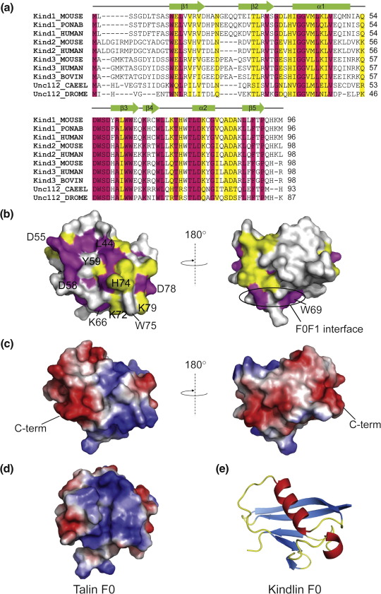
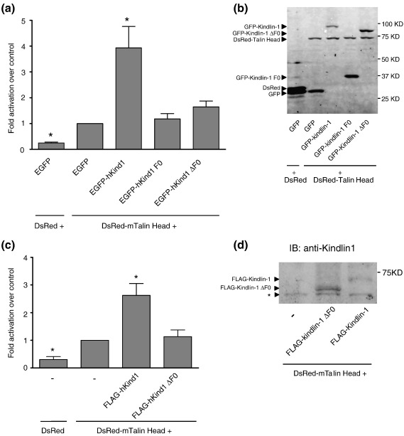
The integrin family of heterodimeric cell adhesion molecules exists in both low- and high-affinity states, and integrin activation requires binding of the talin FERM (four-point-one, ezrin, radixin, moesin) domain to membrane-proximal sequences in the beta-integrin cytoplasmic domain. However, it has recently become apparent that the kindlin family of FERM domain proteins is also essential for talin-induced integrin activation. FERM domains are typically composed of F1, F2, and F3 domains, but the talin FERM domain is atypical in that it contains a large insert in F1 and is preceded by a previously unrecognized domain, F0. Initial sequence alignments showed that the kindlin FERM domain was most similar to the talin FERM domain, but the homology appeared to be restricted to the F2 and F3 domains. Based on a detailed characterization of the talin FERM domain, we have reinvestigated the sequence relationship with kindlins and now show that kindlins do indeed contain the same domain structure as the talin FERM domain. However, the kindlin F1 domain contains an even larger insert than that in talin F1 that disrupts the sequence alignment. The insert, which varies in length between different kindlins, is not conserved and, as in talin, is largely unstructured. We have determined the structure of the kindlin-1 F0 domain by NMR, which shows that it adopts the same ubiquitin-like fold as the talin F0 and F1 domains. Comparison of the kindlin-1 and talin F0 domains identifies the probable interface with the kindlin-1 F1 domain. Potential sites of interaction of kindlin F0 with other proteins are discussed, including sites that differ between kindlin-1, kindlin-2, and kindlin-3. We also demonstrate that F0 is required for the ability of kindlin-1 to support talin-induced alphaIIbbeta3 integrin activation and for the localization of kindlin-1 to focal adhesions.


Figures

Fig. 1
Fig. 1
Comparison of the domain structure of kindlins and the talin head. (a) Schematic diagram of the domain structure of kindlins and talin. The individual domains—F1, F2, and F3—that make up a canonical FERM domain are shown in green, orange, and blue, respectively; the F0 domain is shown in red. The kindlin PH domain is indicated by a black box. Unstructured regions including the F1–loop region are shown in white. The position of the long C-terminal talin rod is indicated. The horizontal scale in both schematics is the same. (b) The primary sequences of mouse kindlin-1 and talin-1 FERM domains were aligned by T-Coffee. The same color scheme is used as in (a). The sequence of the F1 insert and the kindlin PH domain are not included in the alignment. The six tryptophan residues in kindlin-1 F0 are shown in bold. The overall similarity between talin-1 and kindlin-1 is relatively low (28%), due largely to the inclusion of the kindlin PH domain and the insert in F1. The similarity between the individual F0, F1, F2, and F3 domains is much higher (36–55%).
Fig. 2
Fig. 2
The solution structure of the F0 domain of kindlin-1. (a) 1H–15N HSQC spectrum of residues 1–96. (b) Superposition of the 20 lowest-energy solution structures of kindlin-1 F0. (c) Ribbon view of a representative low-energy structure of kindlin-1 F0 showing the β-grasp fold; secondary-structure elements are indicated. (d) The closest structural homologue of F0, ubiquitin (PDB ID: 2GBM), oriented in the same way as kindlin-1 F0 in (c), to show their structural similarity. (e) Ribbon view of kindlin F0 as in (c) with the six tryptophan residues shown in green and the positive charges surrounding W75 shown in purple. (f) The F0 domain of talin-1 showing the remarkable structural similarity between the two F0 domains.
Fig. 3
Fig. 3
Conservation of the kindlin F0 domain. (a) Sequence alignment of mouse kindlin-1 residues 1–96 with the corresponding regions of other kindlins. Magenta, invariant residues; yellow, residues that are highly conserved. The secondary-structure elements are shown above the alignments. The sequences used are as follows: kindlin-1 of mouse (P59113), orangutan (Q5R8M5), and human (Q9BQL6); kindlin-2 of mouse (Q8CIB5) and human (Q96AC1); kindlin-3 of mouse (Q8K1B8), human (Q86UX7), and cow (Q32LP0); Unc-112 of C.elegans (Q18685) and Drosophila (Q9VZ13). (b) Surface representation of the kindlin-1 F0 domain showing the position of the conserved residues identified in (a). A conserved surface comprising helix 1 and the loop between helix 1 and strand 3 is present in all kindlin isoforms. (c) Electrostatic surface of kindlin-1 F0. The left panel is oriented as in (b) and (e). The flexible N-terminus (residues 1–11) is not shown. (d) Electrostatic surface of the talin-1 F0 domain—the orientation is the same as that for kindlin-1 F0 shown in the left panels in (b) and (c). (e) Ribbon representation of kindlin-1 F0 showing the orientation of the protein in the left panels in (b) and (c).
Fig. 4
Fig. 4
The kindlin F0 domain is important for kindlin-1-mediated αIIbβ3 integrin activation. CHO cells stably expressing αIIbβ3 integrin were co-transfected with DsRed or DsRed-tagged talin head (1–433) and GFP or GFP-tagged kindlin-1 cDNAs as indicated. (a) Activation indices of αIIbβ3 integrin from co-expressing cells with similar fluorescence of GFP and DsRed tags were calculated and normalized for integrin expression (see Materials and Methods). The results represent the means ± standard error (n ≥ 3). Results that are significantly different from GFP + DsRed talin head (p < 0.05) are indicated (*). (b) Total lysates from double transfected CHO cells were separated by SDS-PAGE and analyzed by immunoblot for tagged proteins using specific anti-GFP (Rockland), anti-DsRed (Santa Cruz), and Alexa 680 Fluor-coupled anti-goat (Invitrogen) antibodies. (c) αIIbβ3-expressing CHO cells were transfected as above but FLAG-tagged kindlin-1 or kindlin-1ΔF0 was substituted for the GFP-tagged versions. Transfected cells were gated based on equal DsRed fluorescence, and activation indices were calculated and normalized for integrin expression as above. Activation indices were expressed relative to DsRed talin alone and represent means ± standard error (n ≥ 3). (d) Total cell lysates from cells transfected in (c) were separated by SDS-PAGE and probed for recombinant kindlin proteins. CHO cells do not contain detectable levels of endogenous kindlin-1—the nonspecific band (*) migrates faster than that expected for intact kindlin-1.
Fig. 5
Fig. 5
The kindlin-1 F0 domain is required for targeting to FAs. Images of CHO cells stably expressing αIIbβ3 integrin transiently transfected with GFP-tagged kindlin-1 wild-type, F0, and ΔF0 expression constructs after 4 h growth on fibrinogen-coated coverslips. GFP-kindlin-1 co-localizes with endogenous vinculin at FAs (white arrows). Neither GFP-kindlin-1 F0 nor GFP-kindlin-1ΔF0 clusters in vinculin-rich FAs.

References

    1. Aszodi A., Legate K.R., Nakchbandi I., Fassler R. What mouse mutants teach us about extracellular matrix function. Annu. Rev. Cell Dev. Biol. 2006;22:591–621. - PubMed
    1. Harburger D.S., Calderwood D.A. Integrin signalling at a glance. J. Cell Sci. 2009;122:159–163. - PMC - PubMed
    1. Legate K.R., Fassler R. Mechanisms that regulate adaptor binding to β-integrin cytoplasmic tails. J. Cell Sci. 2009;122:187–198. - PubMed
    1. Legate K.R., Montanez E., Kudlacek O., Fassler R. ILK, PINCH and parvin: the tIPP of integrin signalling. Nat. Rev., Mol. Cell Biol. 2006;7:20–31. - PubMed
    1. Zaidel-Bar R., Itzkovitz S., Ma'ayan A., Iyengar R., Geiger B. Functional atlas of the integrin adhesome. Nat. Cell Biol. 2007;9:858–867. - PMC - PubMed

Publication types

MeSH terms

Associated data