. 2009 Oct;85(4):482-92.
doi: 10.1016/j.ajhg.2009.09.005.

Familial hemophagocytic lymphohistiocytosis type 5 (FHL-5) is caused by mutations in Munc18-2 and impaired binding to syntaxin 11

Affiliations

Familial hemophagocytic lymphohistiocytosis type 5 (FHL-5) is caused by mutations in Munc18-2 and impaired binding to syntaxin 11

Udo zur Stadt et al. Am J Hum Genet. 2009 Oct.

Abstract

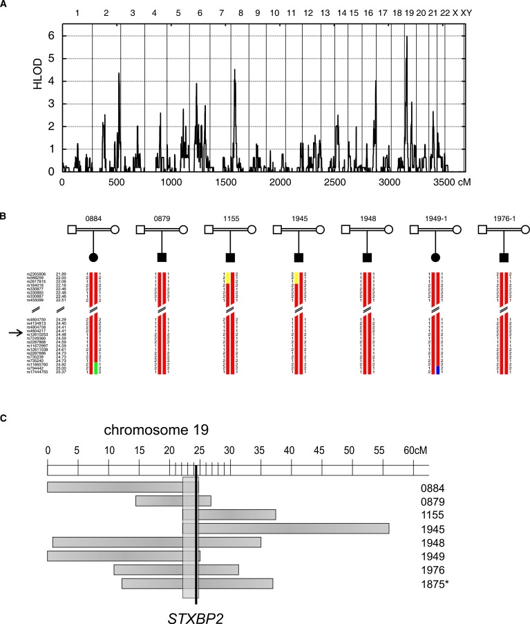
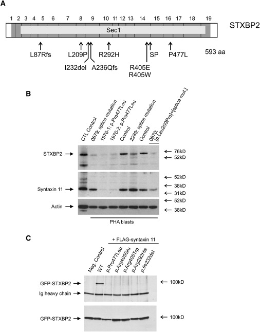
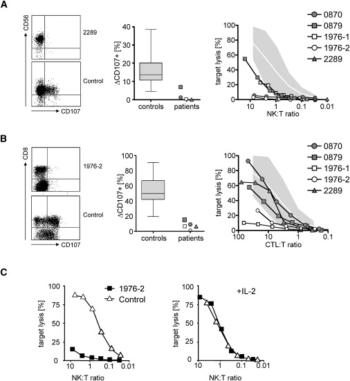
Rapid intracellular transport and secretion of cytotoxic granules through the immunological synapse requires a balanced interaction of several proteins. Disturbance of this highly regulated process underlies familial hemophagocytic lymphohistiocytosis (FHL), a genetically heterogeneous autosomal-recessive disorder characterized by a severe hyperinflammatory phenotype. Here, we have assigned FHL-5 to a 1 Mb region on chromosome 19p by using high-resolution SNP genotyping in eight unrelated FHL patients from consanguineous families. Subsequently, we found nine different mutations, either truncating or missense, in STXBP2 in twelve patients from Turkey, Saudi Arabia, and Central Europe. STXBP2 encodes syntaxin binding protein 2 (Munc18-2), involved in the regulation of vesicle transport to the plasma membrane. We have identified syntaxin 11, a SNARE protein mutated in FHL-4, as an interaction partner of STXBP2. This interaction is eliminated by the missense mutations found in our FHL-5 patients, which leads to a decreased stability of both proteins, as shown in patient lymphocytes. Activity of natural killer and cytotoxic T cells was markedly reduced or absent, as determined by CD107 degranulation. Our findings thus identify a key role for STXBP2 in lytic granule exocytosis.


Figures

Figure 1
Figure 1
Identification of Mutations in STXBP2 Underlying FHL Type 5 (A) A genome-wide linkage scan was performed in 15 single patients with FHL from consanguineous families. LOD scores assuming locus heterogeneity (HLOD scores) were plotted for all autosomes, corresponding to approximately 3500 cM in total length, and revealed a locus on chromosome 19 with a maximum LOD score of 5.9. (B) SNP genotypes of seven FHL patients with homozygosity around STXBP2. Names of markers and their genetic localization are shown on the left. STXBP2 is located between rs4804217 and rs12610253. (C) Eight patients showed overlapping regions of homozygosity on chromosome 19 (horizontal boxes). The minimal region (vertical box) was 1 Mb in length and contained the gene STXBP2. Pedigree 1875 (asterisk) was mapped independently and is not included in the LOD score analysis summarized in (A).
Figure 2
Figure 2
Structure of STXBP2, Protein Stability, and Interaction (A) STXBP2 consists of 19 exons and includes 593 codons (dark gray). It encodes syntaxin binding protein 2 (STXBP2), which contains a large Sec1 domain (light gray). We identified nine different mutations distributed throughout the gene. SP, splice mutation c.1247-1G>C. (B) Lysates from PHA blasts obtained from FHL-5 patients and control persons were analyzed for presence of STXBP2 and syntaxin 11. Patients showed reduced amounts of STXBP2. Moreover, patients with the homozygous STXBP2 missense mutation p.Pro477Leu (1976-1, 1976-2) showed clearly decreased amounts of syntaxin 11. Patient 0870, compound heterozygous for the splice-site mutation c.1247-1G>C and the missense mutation p.Leu209Pro, revealed an intermediate pattern. (C) Coimmunoprecipitation demonstrated shared binding sites between STXBP2 and syntaxin 11. The ability to bind to syntaxin 11 is lost in all STXBP2 mutants with missense mutations and the one-codon deletion identified in our patients with FHL-5 (upper panel). All of the STXBP2 mutants were evenly detectable (lower panel).
Figure 3
Figure 3
Expression of STXBP2 and STX11 Relative quantification of STXBP2 and STX11 in various human tissues by real-time PCR. Expression of both STXBP2 and STX11 is similar and particularly high in NK cells, T cells, monocytes, and other hematopoietic cells. Quantification was done with the use of ACTB, GAPDH, TFRC, and 18S rRNA as references ± SD.
Figure 4
Figure 4
NK and T Cell Degranulation and Cytotoxicity of Patients with Mutations in STXBP2 (A and B) NK cell degranulation was examined by flow cytometry with the use of antibodies against CD107 after stimulation of PBMC with K562 cells (A). Plots are gated on CD3 NKp46+ cells (left panel). CTL degranulation was examined by flow cytometry with the use of antibodies against CD107 after stimulation with anti-CD3/anti-CD28 microbeads (B). Plots are gated on CD3+ cells. The middle panels show the percentage of CD107+ expression on stimulated NK cells and CTLs, respectively, of patients and 30 healthy controls (box, mean and 25%–75% percentile; whiskers, maximum and minimum value observed). The right panels show NK cell cytotoxicity against K562 target cells and CTL cytotoxicity against L1210 target cells, respectively. The shaded areas indicate the mean percentage of target cell lysis ± two standard deviations of 30 healthy controls. The NK:target and CTL:target ratios were determined by flow cytometry. (C) Reconstitution of NK cell cytotoxicity after in vitro stimulation with IL-2. Left panel: Cytotoxicity of PBMC of patient 1976-2 and a healthy control person were assessed against K562 target cells. The NK cell to target ratio was determined by flow cytometry. Right panel: PBMC were cultured for 7 days in medium supplemented with 100 U/ml recombinant IL-2 before analysis of cytotoxicity on K562 target cells.

References

    1. Henter J.I., Samuelsson-Horne A., Arico M., Egeler R.M., Elinder G., Filipovich A.H., Gadner H., Imashuku S., Komp D., Ladisch S. Treatment of hemophagocytic lymphohistiocytosis with HLH-94 immunochemotherapy and bone marrow transplantation. Blood. 2002;100:2367–2373. - PubMed
    1. Stephan J.L., Donadieu J., Ledeist F., Blanche S., Griscelli C., Fischer A. Treatment of familial hemophagocytic lymphohistiocytosis with antithymocyte globulins, steroids, and cyclosporin A. Blood. 1993;82:2319–2323. - PubMed
    1. Arico M., Janka G., Fischer A., Henter J.I., Blanche S., Elinder G., Martinetti M., Rusca M.P. Hemophagocytic lymphohistiocytosis. Report of 122 children from the International Registry. FHL Study Group of the Histiocyte Society. Leukemia. 1996;10:197–203. - PubMed
    1. Janka G.E. Familial hemophagocytic lymphohistiocytosis. Eur. J. Pediatr. 1983;140:221–230. - PubMed
    1. Ohadi M., Lalloz M.R., Sham P., Zhao J., Dearlove A.M., Shiach C., Kinsey S., Rhodes M., Layton D.M. Localization of a gene for familial hemophagocytic lymphohistiocytosis at chromosome 9q21.3–22 by homozygosity mapping. Am. J. Hum. Genet. 1999;64:165–171. - PMC - PubMed

Publication types