. 2009 Oct 13;106(41):17528-33.
doi: 10.1073/pnas.0905523106. Epub 2009 Sep 29.

SH3TC2/KIAA1985 protein is required for proper myelination and the integrity of the node of Ranvier in the peripheral nervous system

Affiliations

SH3TC2/KIAA1985 protein is required for proper myelination and the integrity of the node of Ranvier in the peripheral nervous system

Estelle Arnaud et al. Proc Natl Acad Sci U S A. .

Erratum in

  • Proc Natl Acad Sci U S A. 2010 Aug 24;107(34):15305. Kleine, Henning [added]; Luscher, Bernhard [added]

Abstract

Charcot-Marie-Tooth disease type 4C (CMT4C) is an early-onset, autosomal recessive form of demyelinating neuropathy. The clinical manifestations include progressive scoliosis, delayed age of walking, muscular atrophy, distal weakness, and reduced nerve conduction velocity. The gene mutated in CMT4C disease, SH3TC2/KIAA1985, was recently identified; however, the function of the protein it encodes remains unknown. We have generated knockout mice where the first exon of the Sh3tc2 gene is replaced with an enhanced GFP cassette. The Sh3tc2(DeltaEx1/DeltaEx1) knockout animals develop progressive peripheral neuropathy manifested by decreased motor and sensory nerve conduction velocity and hypomyelination. We show that Sh3tc2 is specifically expressed in Schwann cells and localizes to the plasma membrane and to the perinuclear endocytic recycling compartment, concordant with its possible function in myelination and/or in regions of axoglial interactions. Concomitantly, transcriptional profiling performed on the endoneurial compartment of peripheral nerves isolated from control and Sh3tc2(DeltaEx1/DeltaEx1) animals uncovered changes in transcripts encoding genes involved in myelination and cell adhesion. Finally, detailed analyses of the structures composed of compact and noncompact myelin in the peripheral nerve of Sh3tc2(DeltaEx1/DeltaEx1) animals revealed abnormal organization of the node of Ranvier, a phenotype that we confirmed in CMT4C patient nerve biopsies. The generated Sh3tc2 knockout mice thus present a reliable model of CMT4C neuropathy that was instrumental in establishing a role for Sh3tc2 in myelination and in the integrity of the node of Ranvier, a morphological phenotype that can be used as an additional CMT4C diagnostic marker.


Conflict of interest statement

The authors declare no conflict of interest.

Figures

Fig. 1.
Fig. 1.
Sh3tc2ΔEx1Ex1 mice develop a peripheral neuropathy. (A) Schematic diagram showing the targeting vector containing a 5′ homology arm up to the ATG start site, the eGFP and LoxP-neo-LoxP cassettes necessary for monitoring of the eGFP expression in vivo and Geneticin (G418) selection in ES cells, respectively, and a shorter 3′ homology arm. After homologous recombination in embryonic stem cells, exon 1 (Ex1) of Sh3tc2 gene was eliminated. The neo cassette was removed by crossing the resulting mice with a nestin-Cre deleter strain. Primers used for the ES cell screening (5F and 5R, and 3F and 3R), the mouse genotyping (F, Rwt, and Rmt), and the validation of the neo gene deletion (nF and nR) are indicated by arrows. (B) Amplification of GFP signal using anti-GFP antibody on sciatic nerve cross-cryosections. Typical croissant-shaped staining assessed the Schwann cell specificity of the Sh3tc2 promoter in the homozygous knockout nerve (Upper). No signal was detected in the Sh3tc2+/+ nerves (Lower). (C and D) Nerve conduction velocity of Sh3tc2ΔEx1Ex1 and control mice (n = 4) during their first year of life. At 4 weeks, motor [MNCV, (C)] and sensory [SNCV, (D)] nerve conduction velocities were already significantly slowed down in Sh3tc2ΔEx1Ex1 mice.
Fig. 2.
Fig. 2.
Hypomyelination of sciatic nerve of Sh3tc2ΔEx1Ex1 mice. (A) Electron micrographs of sciatic nerves isolated from wild-type and Sh3tc2ΔEx1Ex1 mice at P56 and at 1 year of age (P365). Although reduced myelin sheath thickness is clearly visible at both stages, the axons themselves remain preserved, even in 1-year-old Sh3tc2ΔEx1Ex1 animals. (B) The scatter plots display myelin thicknesses of individual axons as a function of their respective diameters determined at P10 (n = 4), P23 (n = 4), P56 (n = 2), and P365 (n = 3). Each point corresponds to one fiber (gray points, Sh3tc2ΔEx1Ex1; black points, Sh3tc2+/+). Even at 1 year of age, the myelin in Sh3tc2ΔEx1Ex1 animals did not reach the mature thickness.
Fig. 3.
Fig. 3.
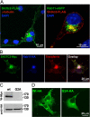
Subcellular localization of Sh3tc2 at the plasma membrane and endocytic recycling compartment. (A) The mouse Schwann cell line (MSC80) cells were transfected with Sh3tc2-FLAG (Left) and with Sh3tc2-FLAG in combination with Rab11-eGFP (Right). Transfected cells were immunostained with anti-FLAG antibody and γ-tubulin and costained with DAPI to visualize the nuclei. Sh3tc2 localized at the membrane surface and in a cytoplasmic compartment near the centrosome (Left, arrow). This compartment is the endocytic recycling compartment, as shown by the colocalization of Sh3tc2-FLAG with Rab11-eGFP (Right). (B) Transferrin (red) was internalized by COS-7 cells that were transfected with Rab11-HA (blue) and SH3TC2-Myc (green). The overlay shows colocalization of the three molecules in the endocytic recycling compartment. (C) Autoradiography after in vitro myristoylation assay in COS-7 cells using [3H]myristic acid (3H-Myr) showed that wild-type SH3TC2 (wt) can be myristoylated at the N terminus, whereas myristoylation is lost if the N-terminal motif is mutated (G2A). (Lower) A Western blot documenting equal expression of mutant and wild-type SH3TC2-HA. (D) When transfected into COS-7 cells, mutation of the SH3TC2 myristoylation site (G2A-HA) led to diffuse cytoplasmic staining and loss of the plasma membrane localization, whereas the localization at the recycling endosome was partially preserved.
Fig. 4.
Fig. 4.
Transcriptional changes in Sh3tc2ΔEx1Ex1 sciatic nerve endoneurium at P28. (A) Variation in the level of transcription of genes involved in myelination. Microarray data are represented as a fold change (positive values represent genes overexpressed and negative values genes underexpressed in Sh3tc2ΔEx1Ex1 mice). Scip, Krox24, and Krox20 encode transcription factors involved in initiation and progression of myelination. Mobp, Mbp, Mpz, Mag, Prx, Pmp22, and Cx32 genes encode myelination-related proteins. (B) Synchronous down-regulation of expression of transcripts encoding enzymes involved in cholesterol synthesis. (C) Representation of the distribution of different functional categories obtained after annotation of genes varying more than 2-fold (P < 0.05) between Sh3tc2ΔEx1Ex1 and control samples. The number of genes in each category is depicted as a fraction of the total number of regulated genes and is also indicated in each fraction of the pie. Some genes were represented by multiple array probes listed in Table S1. Genes with unknown function and biological categories represented by fewer than three genes are not represented.
Fig. 5.
Fig. 5.
Nodes of Ranvier are substantially widened in Sh3tc2ΔEx1Ex1 mice and CMT4C patients' sciatic nerves. (A) Nile red (Nr) staining of myelin of single teased fibers revealed larger nodes of Ranvier (arrowheads) between consecutive myelin sheaths in 5-month-old mutant (Sh3tc2ΔEx1Ex1) compared with control (Sh3tc2+/+) nerve fibers. DAPI staining (blue) enables the visualization of Schwann cell nuclei. (B) Immunostaining of the nodes on teased fibers isolated from 1-year-old animals. Caspr staining confirmed widening of the nodes. Na+ channels were still well clustered at the nodes of Sh3tc2ΔEx1Ex1 fibers, as assessed by Nav staining. Phospho-ERM staining was less intense in mutant nodes, and Kv1.2 distribution at the juxtaparanode was substantially more diffuse in Sh3tc2ΔEx1Ex1 fibers. (C) Longitudinal sections of sciatic nerve isolated from 1-year-old mice analyzed by EM showed wider nodes in Sh3tc2ΔEx1Ex1 axons compared with control samples. Inset shows that the attachment of paranodal loops to the axon, including the presence of transverse bands (arrowheads), is preserved in mutant nerve. Basal lamina covering the node was not affected in Sh3tc2ΔEx1Ex1 animals. (Magnification: Inset, 64,000×.) (D) Nodes of Ranvier observed in human sural nerve biopsy specimens from two CMT4C patients show remarkably similar changes compared with the Sh3tc2ΔEx1Ex1 mouse.

References

    1. Skre H. Genetic and clinical aspects of Charcot-Marie-Tooth's disease. Clin Genet. 1974;6:98–118. - PubMed
    1. Gabreels-Festen AA, Gabreels FJ, Jennekens FG, Joosten EM, Janssen-van Kempen TW. Autosomal recessive form of hereditary motor and sensory neuropathy type I. Neurology. 1992;42:1755–1761. - PubMed
    1. Kessali M, et al. A clinical, electrophysiologic, neuropathologic, and genetic study of two large Algerian families with an autosomal recessive demyelinating form of Charcot-Marie-Tooth disease. Neurology. 1997;48:867–873. - PubMed
    1. Senderek J, et al. Mutations in a gene encoding a novel SH3/TPR domain protein cause autosomal recessive Charcot-Marie-Tooth type 4C neuropathy. Am J Hum Genet. 2003;73:1106–1119. - PMC - PubMed
    1. Houlden H, et al. The phenotype of Charcot-Marie-Tooth disease type 4C due to SH3TC2 mutations and possible predisposition to an inflammatory neuropathy. Neuromuscul Disord. 2009;19:264–269. - PubMed

Publication types

MeSH terms

Substances