. 2009 Oct 6;106(40):17002-6.
doi: 10.1073/pnas.0906095106. Epub 2009 Sep 28.

Crystal structure of Manduca sexta prophenoloxidase provides insights into the mechanism of type 3 copper enzymes

Affiliations

Crystal structure of Manduca sexta prophenoloxidase provides insights into the mechanism of type 3 copper enzymes

Yongchao Li et al. Proc Natl Acad Sci U S A. .

Abstract

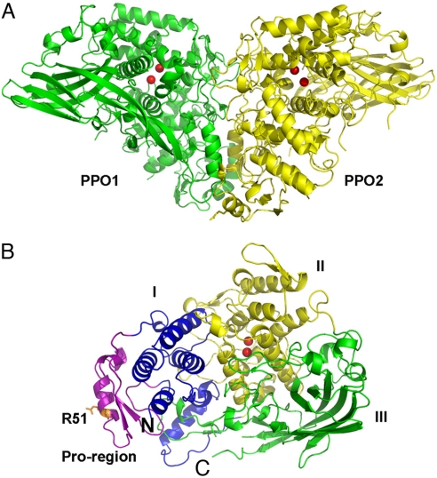
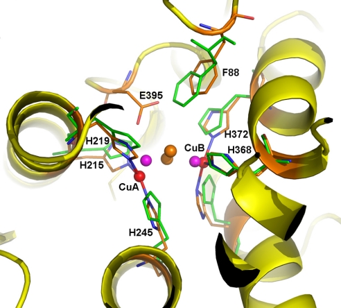
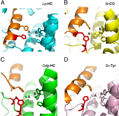
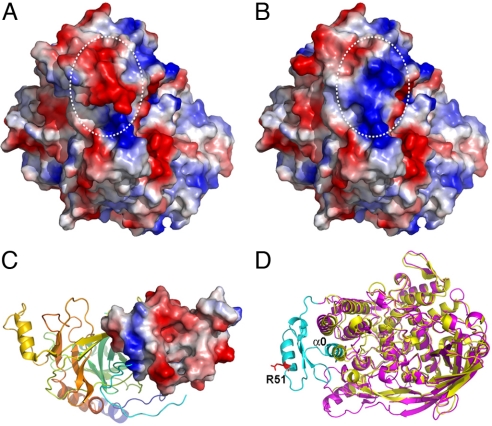
Arthropod phenoloxidase (PO) generates quinones and other toxic compounds to sequester and kill pathogens during innate immune responses. It is also involved in wound healing and other physiological processes. Insect PO is activated from its inactive precursor, prophenoloxidase (PPO), by specific proteolysis via a serine protease cascade. Here, we report the crystal structure of PPO from a lepidopteran insect at a resolution of 1.97 A, which is the initial structure for a PPO from the type 3 copper protein family. Manduca sexta PPO is a heterodimer consisting of 2 homologous polypeptide chains, PPO1 and PPO2. The active site of each subunit contains a canonical type 3 di-nuclear copper center, with each copper ion coordinated with 3 structurally conserved histidines. The acidic residue Glu-395 located at the active site of PPO2 may serve as a general base for deprotonation of monophenolic substrates, which is key to the ortho-hydroxylase activity of PO. The structure provides unique insights into the mechanism by which type 3 copper proteins differ in their enzymatic activities, albeit sharing a common active center. A drastic change in electrostatic surface induced on cleavage at Arg-51 allows us to propose a model for localized PPO activation in insects.


Conflict of interest statement

The authors declare no conflict of interest.

Figures

Fig. 1.
Fig. 1.
Overall structure of M. sexta PPO. (A) The heterodimeric PPO is formed in a back-to-back mode. PPO1 and PPO2 are shown in green and yellow, respectively. (B) Domains of PPO2 are colored as follows: pro-region, purple; domain I, blue; domain II, yellow; domain III, green. The di-copper atoms are located in domain II and are shown as red spheres. The proteolytic site R51 residue is shown in the stick. The amino-terminus and carboxyl-terminus of PPO2 are indicated as N and C, respectively.
Fig. 2.
Fig. 2.
Di-copper center in M. sexta PPO2. The active site of PPO2 can be superimposed well with that of oxygenated Limulus polyphemus hemocyanin (Lp-HC, PDB ID code 1OXY). The secondary structures of PPO2 are shown in the ribbon and colored in yellow. The 6 copper-coordinating His ligands are shown as sticks, with those from Lp-HC colored green. The di-copper atoms are shown as spheres: PPO2, red; Lp-HC, purple. The peroxide ion in Lp-HC is shown as brown spheres. Notice the unique E395 in PPO2, which is located near the substrate place holder F88. E395 could be a base for phenol deprotonation, which is key to the ortho-phenol hydroxylation activity of PPO.
Fig. 3.
Fig. 3.
Different histidine coordination at CuA site. The active sites of 4 representative type 3 copper proteins are shown in the ribbons. Limulus polyphemus hemocyanin (Lp-HC, PDB ID code 1oxy), cyan (A); sweet potato Ipomoea batatas catechol oxidase (Ib-CO, PDB ID code 1bug), yellow (B); Octopus dofleini hemocyanin (Odg-HC, PDB ID code 1js8), green (C); and Streptomyces castaneoglobisporus tyrosinase (Sc-Tyr, PDB ID code 1wx5), salmon (D). The di-copper atoms and their 6-histidine ligands are shown in spheres and sticks, respectively. The unique histidine ligands at CuA sites are colored in red for comparison. The structurally conserved α-helix, where a second histidine locates, is colored in orange for reference in each structure. In Lp-HC, the histidine in comparison is located on the same α-helix where another histidine ligand resides. Notice that the thioether bond in Ib-CO is formed between 2 secondary structures, tethering the histidine residue at the CuA site, which resembles the CuA coordination in Lp-HC. In comparison, the equivalent histidine residues in both Odg-HC and Sc-Tyr are flexible. The thioether bond in Odg-HC is formed within the flexible loop, whereas that in Sc-Tyr does not form any thioether bonds.
Fig. 4.
Fig. 4.
Electrostatic surface potential switching of M. sexta PPO2 on cleavage. The negatively and positively charged surfaces are colored in red and blue, respectively. (A) The intact PPO2 structure. (B) Computer-modeled PPO2 structure after simulated cleavage at R51 and removal of the α-helix where it locates. Notice the oppositely charged surface after cleavage as indicated in white circles. (C) Crystal structure of an SPH from H. diomphalia (PDB ID code 2B9L). The clip domain is shown as an electropotential surface, whereas the rest of the protein is shown in the ribbon. Notice the predominantly negative-charged surface of the clip domain. (D) Superimposition of the structures of PPO2 (purple) and Lp-HC (PDB ID code 1OXY, yellow). The N-terminus of PPO2 flanking the proteolytic site R51 (shown in the red stick) is colored in cyan. Notice that α0 (–16) of PPO2 superimposes well with one of the conserved α-helices in domain I of Lp-HC. Therefore, α0 is considered as part of domain I in PPO2, which may still be associated with the rest of the PO structure even after the proteolytic cleavage.

References

    1. Ashida M, Brey PT. Recent advances on the research of the insect prophenoloxidase cascade. In: Brey PT, Hultmark D, editors. Molecular Mechanisms of Immune Response in Insects. London: Chapman & Hall; 1998. pp. 135–172.
    1. Kanost MR, Gorman MJ. Phenoloxidases in insect immunity. In: Beckage NE, editor. Insect Immunity. New York: Academic; 2008. pp. 69–96.
    1. Nappi AJ, Christensen BM. Melanogenesis and associated cytotoxic reactions: Applications to insect innate immunity. Insect Biochem Mol Biol. 2005;35(5):443–459. - PubMed
    1. Gillespie JP, Kanost MR, Trenczek T. Biological mediators of insect immunity. Annu Rev Entomol. 1997;42:611–643. - PubMed
    1. Lavine MD, Strand MR. Insect hemocytes and their role in immunity. Insect Biochem Mol Biol. 2002;32:1295–1309. - PubMed

Publication types

MeSH terms

Associated data

LinkOut - more resources