. 2009 Oct 6;106(40):17061-6.
doi: 10.1073/pnas.0906823106. Epub 2009 Sep 14.

A wound-induced Wnt expression program controls planarian regeneration polarity

Affiliations

A wound-induced Wnt expression program controls planarian regeneration polarity

Christian P Petersen et al. Proc Natl Acad Sci U S A. .

Abstract

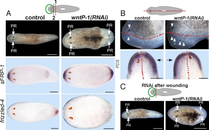
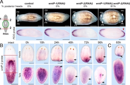
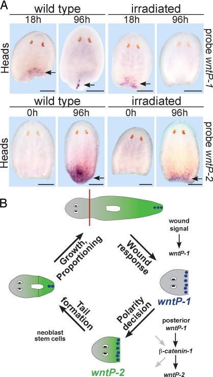
Regeneration requires specification of the identity of new tissues to be made. Whether this process relies only on intrinsic regulative properties of regenerating tissues or whether wound signaling provides input into tissue repatterning is not known. The head-versus-tail regeneration polarity decision in planarians, which requires Wnt signaling, provides a paradigm to study the process of tissue identity specification during regeneration. The Smed-wntP-1 gene is required for regeneration polarity and is expressed at the posterior pole of intact animals. Surprisingly, wntP-1 was expressed at both anterior- and posterior-facing wounds rapidly after wounding. wntP-1 expression was induced by all types of wounds examined, regardless of whether wounding prompted tail regeneration. Regeneration polarity was found to require new expression of wntP-1. Inhibition of the wntP-2 gene enhanced the polarity phenotype due to wntP-1 inhibition, with new expression of wntP-2 in regeneration occurring subsequent to expression of wntP-1 and localized only to posterior-facing wounds. New expression of wntP-2 required wound-induced wntP-1. Finally, wntP-1 and wntP-2 expression changes occurred even in the absence of neoblast stem cells, which are required for regeneration, suggesting that the role of these genes in polarity is independent of and instructive for tail formation. These data indicate that wound-induced input is involved in resetting the normal polarized features of the body axis during regeneration.


Conflict of interest statement

The authors declare no conflict of interest.

Figures

Fig. 1.
Fig. 1.
wntP-1 is expressed at both anterior- and posterior-facing wounds during regeneration. (A–E) wntP-1 in situ hybridizations in intact animals (A) and regenerating head (B), trunk (C and D), and tail fragments (E) at time points (h) after surgery. Brackets, magnified regions at posterior- (B and D) or anterior- (C and E) facing wounds. (F–J) Diagrams depict surgeries. In situ hybridizations with wntP-1 riboprobe at time points after tissue slice (F), hole puncture (G), tissue triangle removal and tail amputation (H), oblique amputation (I), and lateral amputation (J). Anterior, top (A) or left (B–J). (A–E) Dorsal view (A and D, 48 h; B–D, 72 h); other panels, ventral view. (F–J) D, dorsal view; V, ventral view; arrows, regeneration-specific wntP-1 expression. (Scale bars: B–E, 100; A, F–J, 200 μm.) Panels represent ≥4 of 5 animals probed.
Fig. 2.
Fig. 2.
wntP-1 is required for regeneration polarity and anteroposterior patterning. (A) Freshly amputated head fragments (amputation 1) were injected with control or wntP-1 dsRNA for 3 days, amputated again posteriorly on the third day (amputation 2), and allowed to regenerate for 12 days. (Upper) Control head fragments regenerated normally (100%, n = 35), whereas wntP-1(RNAi) animals regenerated a posterior-facing head (26%, n = 77, three experiments). (Lower) In situ hybridizations of control or wntP-1(RNAi) animals at 12 days of regeneration probed with sFRP-1 and frizzled-4 riboprobes. Images are representatives: sFRP-1-stained wntP-1(RNAi), 6 of 24 animals; other panels ≥7 of 7 animals. (B) Intact animals were injected for 2 days and amputated sagittally the following day. (Upper) Uninjected lateral fragments regenerated normally (100%, n = 46), whereas wntP-1(RNAi) fragments regenerated with supernumerary photoreceptors (40%, n = 42) by 10 days. (Lower) in situ hybridizations of laterally regenerating control and wntP-1(RNAi) fragments using a prostaglandin-D synthetase (PDS) riboprobe. Black arrows, newly regenerated side. Extent of PDS in situ hybridization signal was greater on the regenerated side in wntP-1(RNAi) (3 of 5 animals with extra photoreceptors), but not in control animals (7 of 7 animals). Red line, approximate old/new tissue boundary. (C) Day 13 regenerating head fragments that were injected with indicated dsRNAs 1 h after decapitation. Diagrams: RNAi and surgical strategies. PR, photoreceptor. Anterior, left (A, B Upper, C) or top (B Lower). (Scale bars: 200 μm.)
Fig. 3.
Fig. 3.
wntP-2 acts with wntP-1 to control regeneration polarity. (A) Diagram depicts RNAi and surgical strategy. Freshly amputated trunk fragments were injected with indicated dsRNA for three consecutive days, amputated near the anterior and posterior wound sites again on the third day, and allowed to regenerate 14 days. wntP-1 and wntP-2 individual injections were mixed with equal amounts of control dsRNA. (Upper) Percentage of animals that regenerated a posterior head. (Lower) In situ hybridizations of regenerating trunk fragments with PDS riboprobe. Red arrow, anterior marker expression in posterior blastema of wntP-1(RNAi); wntP-2(RNAi) double RNAi animals. (B) wntP-2 in situ hybridizations of intact animals and regenerating head and tail fragments at time points (h) after amputation. (B) wntP-2 was detected in an apparent posterior-to-anterior gradient, internally at the anterior pharynx end. New expression of wntP-2 was detected at posterior wounds in regenerating head fragments from 24 h and at progressively anterior locations as regeneration proceeded (p, expression at the new pharynx in a tail fragment). Black arrows, new wntP-2 expression during regeneration (heads) or anterior limit of wntP-2 expression (tails). (C) wntP-2 expression was detected in the posterior of head fragments (solid arrow) but not in the anterior of decapitated fragments at 65 h (empty arrow). PR, photoreceptor. Anterior, left (A) or top (B and C). (Scale bars: 200 μm.)
Fig. 4.
Fig. 4.
wntP-2 expression at posterior-facing wounds requires wntP-1. In situ hybridizations with wntP-1 (A) or wntP-2 (B) riboprobes of regenerating head fragments at time points (h), which received control, wntP-1, or β-catenin-1 dsRNA before decapitation. For the 36-h time point, freshly amputated head fragments were injected with dsRNA on two consecutive days, amputated posteriorly the following day, and fixed 36 h later. This strategy resulted in 26% penetrance of posterior head regeneration for wntP-1 inhibition (Fig. 2A). For 0, 18, 42, and 72 h time points, intact animals were injected with dsRNA 48 and 24 h before decapitation, and fixation occurred at the indicated times. This RNAi strategy did not result in regeneration of posterior heads for wntP-1 inhibition (10 of 10 animals) but was sufficient to perturb wntP-2 expression (B). (B) (Left) 0 h control, 0 h wntP-1(RNAi), and 18 h β-catenin-1(RNAi). (Lower Left) Scoring (number similar to that shown in the panel versus total number of animals). wntP-2 expression was absent (3 of 5, 36 h; 4 of 7 42 h; 5 of 7 72 h) or much less abundant than control RNAi (2 of 5, 36 h; 3 of 7, 42 h; 2 of 7, 72 h) after wntP-1 RNAi. Arrows indicate new wntP-1 or wntP-2 expression due to regeneration. Anterior, left. Dorsal view (A, 72 h; B, 0, 18, 72 h); all other panels, ventral view. (Scale bars: 200 μm.)
Fig. 5.
Fig. 5.
Dynamic Wnt expression is stem-cell-independent. (A) In situ hybridizations of wntP-1 and wntP-2 in normal animals (“wild type”) or animals treated with 6,000 rads of γ irradiation 4 days (“irradiated”) before surgical removal of heads, trunks, and tails and fixation at time points (h, hours) after amputation. (Upper) wntP-1 expression was detected at posterior-facing wounds in irradiated head fragments at 18 h but not at 96 h. (Lower) wntP-2 expression was detected in the posterior of 96-h heads. Anterior, top. Arrows, new expression of wntP-1 or wntP-2. Panels represent ≥4 of 5 animals. (Scale bars: 200 μm.) (B) Model of regeneration polarity in head fragments. wntP-1 is induced at any wound. Posterior wntP-1 activates wntP-2 through β-catenin-1, possibly acting in parallel with other genes or resulting in activation of other genes that contribute to the polarity decision (gray arrows). The polarity decision is stem-cell-independent, but tail formation subsequently requires neoblast stem cells. Purple spots, wntP-1 expression. Green gradient, wntP-2 expression.

References

    1. Brockes JP, Kumar A. Comparative aspects of animal regeneration. Annu Rev Cell Dev Biol. 2008;24:525–549. - PubMed
    1. Harrison RG. Experiments on the development of the fore-limb of Amblystoma, a self-differentiating equipotential system. J Exp Zool. 1918;25:413–461.
    1. Reddien PW, Bermange AL, Kicza AM, Sánchez Alvarado A. BMP signaling regulates the dorsal planarian midline and is needed for asymmetric regeneration. Development. 2007;134:4043–4051. - PubMed
    1. Molina MD, Saló E, Cebrià F. The BMP pathway is essential for re-specification and maintenance of the dorsoventral axis in regenerating and intact planarians. Dev Biol. 2007;311:79–94. - PubMed
    1. Whitehead GG, Makino S, Lien CL, Keating MT. fgf20 is essential for initiating zebrafish fin regeneration. Science. 2005;310:1957–1960. - PubMed

Publication types

MeSH terms

LinkOut - more resources