. 2009 Oct 13;106(41):17425-30.
doi: 10.1073/pnas.0909289106. Epub 2009 Oct 1.

Membrane domains and flagellar pocket boundaries are influenced by the cytoskeleton in African trypanosomes

Affiliations

Membrane domains and flagellar pocket boundaries are influenced by the cytoskeleton in African trypanosomes

Catarina Gadelha et al. Proc Natl Acad Sci U S A. .

Abstract

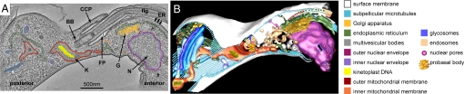
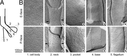
A key feature of immune evasion for African trypanosomes is the functional specialization of their surface membrane in an invagination known as the flagellar pocket (FP), the cell's sole site of endocytosis and exocytosis. The FP membrane is biochemically distinct yet continuous with those of the cell body and the flagellum. The structural features maintaining this individuality are not known, and we lack a clear understanding of how extracellular components gain access to the FP. Here, we have defined domains and boundaries on these surface membranes and identified their association with internal cytoskeletal features. The FP membrane appears largely homogeneous and uniformly involved in endocytosis. However, when endocytosis is blocked, receptor-mediated and fluid-phase endocytic markers accumulate specifically on membrane associated with four specialized microtubules in the FP region. These microtubules traverse a distinct boundary and associate with a channel that connects the FP lumen to the extracellular space, suggesting that the channel is the major transport route into the FP.


Conflict of interest statement

The authors declare no conflict of interest.

Figures

Fig. 1.
Fig. 1.
When endocytosis is blocked by cold treatment of bloodstream-form trypanosomes, markers for endocytosis accumulate specifically at the membrane abutting the 4MT. Representative thin sections of the FP of cells incubated for 15 min on ice with 5-nm gold conjugated to TL or WGA, or unconjugated 5-nm gold (Au). Red bars indicate the positions at which the 4MT pass through the section. Graph shows densities of gold particles on the membrane adjacent to the 4MT or elsewhere (expressed as particles per micrometer of membrane observable in thin section). Bars represent standard errors of the mean.
Fig. 2.
Fig. 2.
Electron tomographic reconstruction used to model an entire FP, surrounding organelles, and cytoskeleton. (A) A single ≈2-nm slice from a 250-nm-thick serial-section 3D reconstruction of the anterior FP region from a late M-phase bloodstream-form trypanosome with some of the structures modeled by segmentation along organelle contours. The FP can be seen clearly near the center of the slice, as can the anterior kinetoplast (K), Golgi (G), and the dividing nucleus (N). The anterior–posterior polarity of the trypanosome cell (defined by its swimming direction) is also indicated. The full reconstruction can be seen in Movie S1. (B) A full 3D segmentation model formed from the serial-section reconstruction by the process shown in A. The relationships between individual structures can now be seen clearly. This segmentation model can also be seen in Movie S2 and Movie S3.
Fig. 3.
Fig. 3.
Clathrin structures seen in tomographic reconstructions of the trypanosome FP. (A and B) Both highly curved pits (A) and also flatter areas (B) can be seen (images are 5-nm tomographic slices from tomograms). Clathrin triskelion assembly is not seen near the 4MT or at the collar. No gross segregation to particular subdomains is observed. (C–E) FP topography from segmentation models of electron tomographic reconstructions of whole FP in cells in G1 (C), nuclear S phase (D), or late mitosis (E). Patches of clathrin assembly are shown in light blue, along with the 4MT (dark blue), the collar (pink), and FAZ maculae (green). The newly identified microtubule associated with the neck region is also shown (red). The total surface area of the FP and the percentage that is coated by clathrin are indicated on the right of each panel. The surface area of some individual clathrin-coated pits are also indicated by white arrows. In D, NFP and OFP indicate new and old FPs, respectively.
Fig. 4.
Fig. 4.
Freeze fracture of the FP membrane. Note that IMPs are homogeneously distributed, with a higher numerical density on the P face than the E face. Invaginated membrane domains, representing sites of endocytosis or exocytosis, are indicated by arrowheads. Platinum/carbon shadow direction is from bottom to top. Insets show FP membrane faces at higher magnification.
Fig. 5.
Fig. 5.
The cell surface, FP, and flagellar membranes have different densities of IMPs and are separated by boundary elements with distinctive patterns of particles. Representative images of IMP density and distribution in the membrane regions illustrated by the diagram in A are shown in B. P faces and E faces of each membrane region are shown, together with views of the neck (boundary between the surface and FP membranes) and the flagellum base (boundary between the FP and flagellum membranes). Arrowheads indicate rows of IMPs at the base of the neck. Black arrows indicate direction of platinum/carbon evaporation. Note that panels are presented to match the orientation of the diagram in A. Magnification bar applies to all frames.
Fig. 6.
Fig. 6.
The 4MT in the neck region are associated with a channel that connects the FP to the extracellular space. Transverse thin-section electron micrographs of the neck and flagellum exit site in cells incubated for 15 min on ice with TL conjugated to 5-nm gold. There is a clear accumulation of gold particles in a gap between the flagellum membrane and the neck membrane directly abutting the 4MT (red arrows). Red bars indicate the positions at which the 4MT pass through the section.

References

    1. Grunfelder CG, et al. Endocytosis of a glycosylphosphatidylinositol-anchored protein via clathrin-coated vesicles, sorting by default in endosomes, and exocytosis via RAB11-positive carriers. Mol Biol Cell. 2003;14:2029–2040. - PMC - PubMed
    1. Ferguson MAJ. The structure, biosynthesis, and functions of glycosylphosphatidylinositol anchors and the contributions of trypanosome research. J Cell Sci. 1999;112:2799–2809. - PubMed
    1. Borst P, Fairlamb AH. Surface receptors and transporters of Trypanosoma brucei. Annu Rev Microbiol. 1998;52:745–778. - PubMed
    1. Vanhollebeke B, et al. A haptoglobin-hemoglobin receptor conveys innate immunity to Trypanosoma brucei in humans. Science. 2008;320:677–681. - PubMed
    1. Overath P, Engstler M. Endocytosis, membrane recycling, and sorting of GPI-anchored proteins: Trypanosoma brucei as a model system. Mol Microbiol. 2004;53:735–744. - PubMed

Publication types

LinkOut - more resources