. 2009 Oct 13;106(41):17564-9.
doi: 10.1073/pnas.0909267106. Epub 2009 Sep 24.

AnkyrinG is required to maintain axo-dendritic polarity in vivo

Affiliations

AnkyrinG is required to maintain axo-dendritic polarity in vivo

Jürgen-Markus Sobotzik et al. Proc Natl Acad Sci U S A. .

Abstract

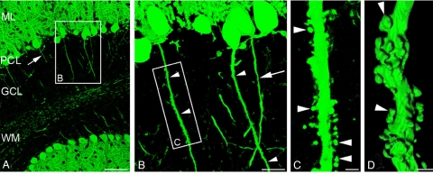
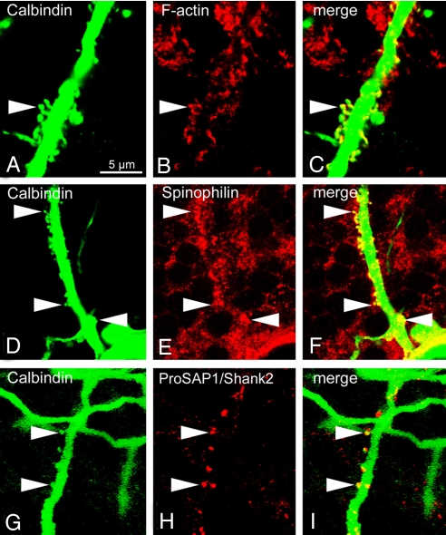
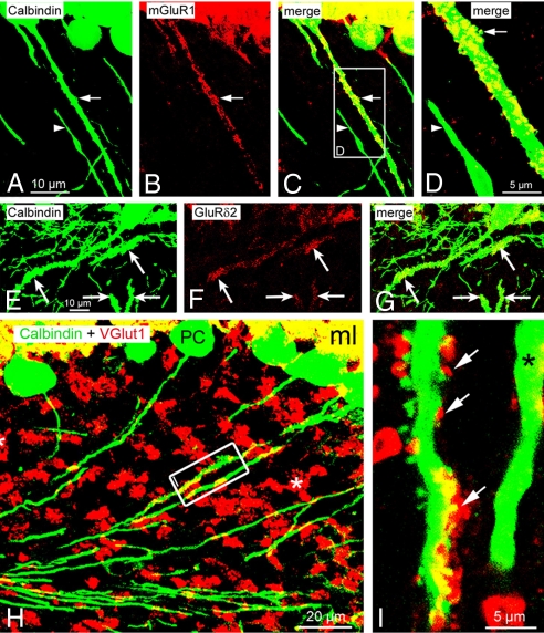
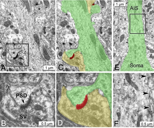
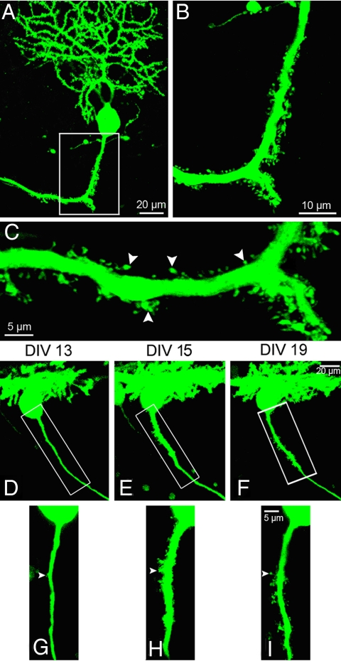
Neurons are highly polarized cells that extend a single axon and several dendrites. Studies with cultured neurons indicate that the proximal portion of the axon, denoted as the axon initial segment (AIS), maintains neuronal polarity in vitro. The membrane-adaptor protein ankyrinG (ankG) is an essential component of the AIS. To determine the relevance of ankG for neuronal polarity in vivo, we studied mice with a cerebellum-specific ankG deficiency. Strikingly, ankG-depleted axons develop protrusions closely resembling dendritic spines. Such axonal spines are enriched with postsynaptic proteins, including ProSAP1/Shank2 and ionotropic and metabotropic glutamate receptors. In addition, immunofluorescence indicated that axonal spines are contacted by presynaptic glutamatergic boutons. For further analysis, double mutants were obtained by crossbreeding ankG(-/-) mice with L7/Purkinje cell-specific promoter 2 (PCP2) mice expressing enhanced green fluorescent protein (EGFP) in Purkinje cells (PCs). This approach allowed precise confocal microscopic mapping of EGFP-positive spiny axons and their subsequent identification at the electron microscopic level. Ultrastructurally, axonal spines contained a typical postsynaptic density and established asymmetric excitatory synapses with presynaptic boutons containing synaptic vesicles. In the shaft of spiny axons, typical ultrastructural features of the AIS, including the membrane-associated dense undercoating and cytoplasmic bundles of microtubules, were absent. Finally, using time-lapse imaging of organotypic cerebellar slice cultures, we demonstrate that nonspiny PC axons of EGFP-positive/ankG(-/-) mice acquire a spiny phenotype within a time range of only 3 days. Collectively, these findings demonstrate that axons of ankG-deficient mice acquire hallmark features of dendrites. AnkG thus is important for maintaining appropriate axo-dendritic polarity in vivo.


Conflict of interest statement

The authors declare no conflict of interest.

Figures

Fig. 1.
Fig. 1.
AnkG-deficiency causes PC axons to develop spines in vivo. (A–C) The morphology of PC in ankG−/− mice is visualized by anti-calbindin D28K labeling. (A) At low magnification, principal features of axo-dendritic polarity are preserved. PC dendrites branch in the molecular layer (ML), PC somata are aligned in the Purkinje cell layer (PCL), and PC axons (arrow) traverse the granule cell layer (GCL) en route to the white matter (WM). (B) Higher magnification of framed area in A. Three PC axons originating from their parental cell body are depicted. Two of them (arrowheads) exhibit a conspicuous spiny appearance. By comparison, the third axon (arrow) shows a smooth surface. (C) Close-up view of framed area in B. The PC axon gives rise to numerous spiny protrusions. These spines have a thread-like neck and a mushroom-like head (arrowheads) and thus resemble dendritic spines. (D) The 3-dimensional morphology of a spiny PC axon is visualized by surface rendering of a confocal Z-stack. (Scale bars: A, 100 μm; B, 20 μm; C and D, 2 μm.)
Fig. 2.
Fig. 2.
Spiny PC axons of ankG−/− mice possess neurochemical features of axons and dendrites. (A–C) Enrichment of filamentous actin in spines of an ankG−/− PC axon (arrowhead). F-actin was labeled by fluorescent phalloidin. (D–F) Enrichment of the dendrite-specific protein spinophilin in spines of an ankG−/− PC axon (arrowheads). (G–I) Highly selective enrichment of the PSD protein ProSAP1/Shank2 in spines of an ankG−/− PC axon (arrowheads). (Scale bar, 5 μm.)
Fig. 3.
Fig. 3.
Features of glutamatergic synapses associated with spines of ankG−/− PC axons. (A–C) Clustering of postsynaptic mGluR1 in a spiny PC axon (arrow). By comparison, a neighboring nonspiny PC axon (arrowhead) is completely devoid of mGluR1. (D) Close-up view of the framed area shown in C. Arrow points to a spine enriched with mGluR1. Arrowhead: nonspiny PC axon devoid of mGluR1. (E–G) Enrichment of postsynaptic GluRδ2 in spiny PC axons (arrows). Nonspiny axons beneath the PC layer belong to the plexus of PC axon collaterals. (H and I) Double labeling for calbindin D28K and VGlut1. (H) Asterisk denotes glutamatergic mossy fiber boutons associated with cerebellar glomeruli. Two PC axons in the framed area are shown at higher magnification in I. (I) Spines of a PC axon are contacted by VGlut1-positive boutons (arrows). A neighboring nonspiny axon (asterisk) is devoid of VGlut1-positive terminals.
Fig. 4.
Fig. 4.
Ultrastructural features of spiny axons in L7-EGFP/ankG−/− double mutants. (A and C) The shaft of an ankG-depleted axon gives rise to mushroom-like spines (asterisks in A). Shades of color in C delineate the contour of a spiny axon (green) and 2 associated presynaptic terminals (yellow). PSDs associated with the axonal spines are shaded in red. See Fig. S3 for confocal scanning microscopy of the same spiny axon. (B and D) Close-up view of the framed area in A. The spine of the ankG-depleted axon is endowed with a PSD and contacted by a presynaptic terminal containing numerous synaptic vesicles (SV). Colors in D correspond to those in C. (E and F) Ultrastructure of the ankG-depleted AIS. E depicts the transition of a PC soma into the AIS of the spiny axon also shown in A–D (contour delineated by green color). (F) Close-up view of the framed area in E. The cytoplasmic membrane of the ankG-depleted AIS (arrowheads) lacks the dense undercoating normally found in the AIS. Likewise, the AIS cytoplasm is devoid of fasciculated microtubules. For comparison with an AIS of a control animal see Fig. S6.
Fig. 5.
Fig. 5.
Identification and time-lapse imaging of spiny axons in cerebellar slice cultures prepared from L7-EGFP/ankG−/− mice. (A–C) EGFP-positive spiny PC axon identified in a cerebellar slice culture (DIV10) of an L7-EGFP/ankG−/− mouse. The spiny appearance of this process (arrowheads) closely resembles that of axons found in cerebellar brain sections of ankG−/− mice (see Fig. 1). (D–I) Confocal time-lapse imaging of a cerebellar slice culture reveals the conversion of a nonspiny into a spiny axon. At DIV13, the selected axon is largely devoid of spines except for a single protrusion (arrowhead). Between DIV13 (D) and DIV15 (E) the conversion into a spiny axon occurs. This conversion into an axo-dendritic hybrid is accompanied by an enlargement of the axonal diameter. (Scale bars: A and D–F, 20 μm; B, 10 μm; C and G–I, 5 μm.)

References

    1. Craig AM, Banker G. Neuronal polarity. Annu Rev Neurosci. 1994;17:267–310. - PubMed
    1. Winckler B, Mellman I. Neuronal polarity: Controlling the sorting and diffusion of membrane components. Neuron. 1999;23:637–640. - PubMed
    1. Horton AC, Ehlers MD. Neuronal polarity and trafficking. Neuron. 2003;40:277–295. - PubMed
    1. Arimura N, Kaibuchi K. Neuronal polarity: From extracellular signals to intracellular mechanisms. Nat Rev Neurosci. 2007;8:194–205. - PubMed
    1. Barnes AP, Solecki D, Polleux F. New insights into the molecular mechanisms specifying neuronal polarity in vivo. Curr Opin Neurobiol. 2008;18:44–52. - PMC - PubMed

Publication types