. 2009 Oct 20;106(42):17729-34.
doi: 10.1073/pnas.0909589106. Epub 2009 Oct 1.

Structure of an integrin alphaIIb beta3 transmembrane-cytoplasmic heterocomplex provides insight into integrin activation

Affiliations

Structure of an integrin alphaIIb beta3 transmembrane-cytoplasmic heterocomplex provides insight into integrin activation

Jun Yang et al. Proc Natl Acad Sci U S A. .

Abstract

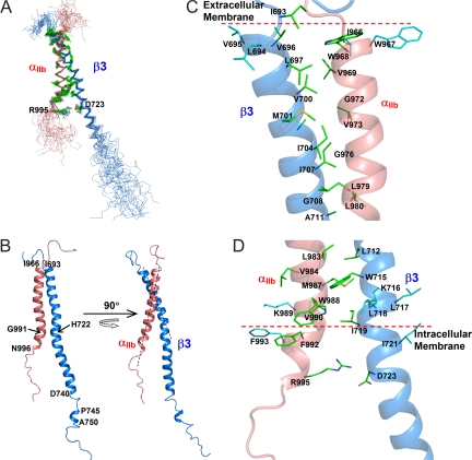
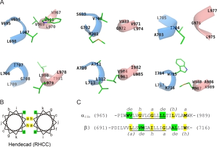
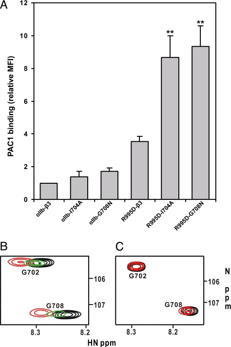
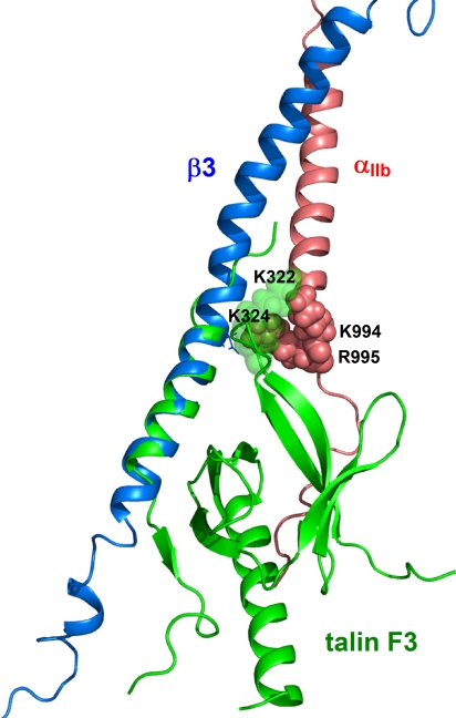
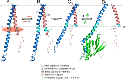
Heterodimeric integrin adhesion receptors regulate diverse biological processes including angiogenesis, thrombosis and wound healing. The transmembrane-cytoplasmic domains (TMCDs) of integrins play a critical role in controlling activation of these receptors via an inside-out signaling mechanism, but the precise structural basis remains elusive. Here, we present the solution structure of integrin alphaIIb beta3 TMCD heterodimer, which reveals a right-handed coiled-coil conformation with 2 helices intertwined throughout the transmembrane region. The helices extend into the cytoplasm and form a clasp that differs significantly from a recently published alphaIIb beta3 TMCD structure. We show that while a point mutation in the clasp interface modestly activates alphaIIb beta3, additional mutations in the transmembrane interface have a synergistic effect, leading to extensive integrin activation. Detailed analyses and structural comparison with previous studies suggest that extensive integrin activation is a highly concerted conformational transition process, which involves transmembrane coiled-coil unwinding that is triggered by the membrane-mediated alteration and disengagement of the membrane-proximal clasp. Our results provide atomic insight into a type I transmembrane receptor heterocomplex and the mechanism of integrin inside-out transmembrane signaling.


Conflict of interest statement

The authors declare no conflict of interest.

Figures

Fig. 1.
Fig. 1.
Structure of integrin αIIbβ3 TMCD heterodimer. (A) Superposition of 20 calculated structures with the lowest energies showing how well the backbone and side chains of the interface containing regions are defined. Notably, the αIIb R995/β3 D723 side chains converge, pointing to each other at short distances and allowing salt bridge formation. (B) Two different views of the entire diagram of the αIIbβ3 TMCD heterodimer. Notice the significant kinks at the transmembrane-cytoplasmic border, which promote formation of the cytoplasmic clasp. (C) Detailed N-terminal half of the interface of the TMCD heterodimer. The side chains, not in the interface but involved in the extracellular membrane embedding, are also shown (colored in cyan). (D). Detailed C-terminal half of the interface. The side chains, not in the interface but involved in the membrane anchoring at the transmembrane-cytoplasmic border, are colored in cyan. The positions of the charged groups of αIIb K989 and β3 K716 (if linearly extended toward the membrane) indicate the TM-CT border (dotted line).
Fig. 2.
Fig. 2.
Coiled coil features of αIIb/β3 TM interface. (A) Cross sections of each turn of both helices with the side-chains of interacting residues shown as sticks. The blue helix is β3 subunit and the red helix is αIIb subunit. (B) Helical wheel projections of a right-handed coiled coil with an 11-residue hendecad repeat (indexed ak). (C) Sequences of the transmembrane domain of αIIb/β3. Residues occupying the a/h indices are colored yellow and de indices are colored green. The underlined regions show the most intensive intermolecular interactions and represent a classical hendecad repeat. The positions of residues with parenthesized indices are slightly shifted in the hendecad repeat extensions.
Fig. 3.
Fig. 3.
Structural and functional effects of CT and TM mutations on integrin activation. (A) Quantitative comparison of the activation of wild type αIIbβ3 vs. mutants containing αIIb R995D, β3 I704A, β3 G708N, and the double mutations αIIb R995D/β3 I704A and αIIb R995D/β3 G708N. The extent of activation was determined from the ratio of PAC1 (activation specific mAb) to 2G12 (αIIbβ3 reactive mAb) binding as measured by FACS. This ratio was assigned a value of 1 for WT, and the activation state of each mutant is compared to WT (34). Results presented are means ± SD from 3 independent experiments. **, P < 0.01 (vs. single mutation) by Student t test. (B) Representative regions of 2D 1H-15N HSQC spectra of 0.1 mM 15N-labeled β3 TMCD in the absence (black) and presence of 0.3 mM WT αIIb TMCD (red) and 0.3 mM αIIb R995D mutant (green) showing that the mutation significantly weakens the αIIb/β3 TMCD association as judged by the substantially reduced chemical shift changes. (C) Representative regions of 2D 1H-15N HSQC spectra of 0.1 mM 15N-labeled β3 TMCD I704A in the absence (black) and presence of 0.4 mM αIIb R995D (red) showing that mutations diminished the αIIb/β3 TMCD association since little chemical shift changes occur.
Fig. 4.
Fig. 4.
Talin disrupts the CT clasp via steric clash and charge-charge repulsion. The β3 membrane-proximal region (bound to talin F3, PDB ID 2h7e) (H722-A737) (35) was superimposed with the same segment in the αIIbβ3 heterodimer, showing how talin F3 K322/K324 may directly interfere with αIIb K994/R995.
Fig. 5.
Fig. 5.
A model for integrin inside-out TM signaling. (A). Step 1: the membrane-proximal clasp is bound to an inhibitor (shaded in red oval) that maintains the integrin at the resting state. (B). Step 2: a cellular signal dissociates the inhibitor from the clasp, leading to an intermediate state of the integrin TMCD heterodimer. (C). Step 3: the cytoplasm-exposed hydrophobic residues (in cyan) upon release of the inhibitor insert or anchors to the membrane, leading to the alteration and membrane-embedding of the clasp (in cyan). (D). Step 4: integrin regulators such as talin (in green) further perturb the clasp, leading to the dissociation of the clasp and subsequent unwinding of the TM coiled-coil. We note that talin could also compete with the inhibitor in Step 1 for binding to the clasp, but other regulators such as migfilin (40) may act synergistically to promote the release of the inhibitor more effectively.

References

    1. Hynes RO. Integrins: Bidirectional, allosteric signaling machines. Cell. 2002;110:673–687. - PubMed
    1. Qin J, Vinogradova O, Plow EF. Integrin bidirectional signaling: A molecular view. PLoS Biol. 2004;2:714–717. - PMC - PubMed
    1. Adair BD, Yeager M. Three-dimensional model of the human platelet integrin alphaIIbbeta 3 based on electron cryomicroscopy and x-ray crystallography. Proc Natl Acad Sci USA. 2002;99:14059–14064. - PMC - PubMed
    1. Schneider D, Engelman DM. Involvement of transmembrane domain interactions in signal transduction by alpha/beta integrins. J Biol Chem. 2004;279:9840–9846. - PubMed
    1. Luo BH, Springer TA, Takagi J. A specific interface between integrin transmembrane helices and affinity for ligand. PLoS Biol. 2004;2:776–786. - PMC - PubMed

Publication types

MeSH terms

Substances

Associated data