. 2009 Sep 15;106(37):15720-5.
doi: 10.1073/pnas.0908450106. Epub 2009 Sep 8.

Feeder-free derivation of induced pluripotent stem cells from adult human adipose stem cells

Affiliations

Feeder-free derivation of induced pluripotent stem cells from adult human adipose stem cells

Ning Sun et al. Proc Natl Acad Sci U S A. .

Abstract

Ectopic expression of transcription factors can reprogram somatic cells to a pluripotent state. However, most of the studies used skin fibroblasts as the starting population for reprogramming, which usually take weeks for expansion from a single biopsy. We show here that induced pluripotent stem (iPS) cells can be generated from adult human adipose stem cells (hASCs) freshly isolated from patients. Furthermore, iPS cells can be readily derived from adult hASCs in a feeder-free condition, thereby eliminating potential variability caused by using feeder cells. hASCs can be safely and readily isolated from adult humans in large quantities without extended time for expansion, are easy to maintain in culture, and therefore represent an ideal autologous source of cells for generating individual-specific iPS cells.


Figures

Fig. 1.
Fig. 1.
Tracking the appearance and growth of an ES cell-like colony by immunostaining the living cells with TRA-1–60 in feeder-free reprogramming. Adult hASCs were seeded on Matrigel-coated surface without MEF feeder cells. The living cells were stained repetitively with TRA-1–60 monoclonal antibodies and AlexaFluor 488 secondary antibodies over the indicated period. Day 18 and 22 images are presented both in fluorescent and phase contrast microscopy. Note that TRA-1–60 expression was specific for the ES cell-like colony. (Scale bar, 100 μm.)
Fig. 2.
Fig. 2.
Characterization of hASC-iPS cells. (A) Immunostaining of hASC-iPS cell colonies with common hES cell markers. The two phase contrast microscopies show a typical hASC-iPS cell colony growing on MEF feeder cells and feeder-free Matrigel surface, respectively. (Scale bars, 100 μm.) (B) Quantitative-PCR analyzing pluripotency gene expression level within hASCs and hASC-iPS cells relative to those in H9 hES cells. iPS_I1–4 denotes iPS cell line #4 derived from individual 1. (C) Bisulphite pyrosequencing measuring methylation status within the promoter region of Oct4 and Nanog genes in H9 hES cells, hASC-iPS cells, hASCs, and IMR90 cells. TSS, transcription start site. (D) Microarray data comparing global gene expression profiles of hASCs, hASC-iPS cells, and hES cells. Upper panel, heat map and hierarchical clustering analysis by Pearson correlation showing hASC-iPS cells are similar to hES cells and distinct from hASCs. Lower panel, scatter plots comparing global gene expression patterns between hASCs, hASC-iPS cells, and hES cells. Highlighted are the pluripotency genes Oct4, Sox2, and Nanog (red arrows). The green diagonal lines indicated linear equivalent and 5-fold changes in gene expression levels between paired samples.
Fig. 3.
Fig. 3.
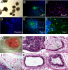
hASC-iPS cells are pluripotent. (A) hASC-iPS cells form EBs and can differentiate to cells of (B and C) endoderm [α-fetoprotein (AFP) and sox17], (D) mesoderm (desmin), and (E and F) ectoderm (Tuj-1 positive motor neurons) lineages. (F) Represents the enlarged view of the boxed area in (E). (Scale bars, 100 μm.) (GL) Upon injection into nude mice, hASC-iPS cells form (G) teratoma in vivo, which contains tissues of all three embryonic germ layers, such as (H) neural epithelium (ectoderm), (I) smooth muscle (arrow) and (J) adipose tissue (mesoderm), and (K) gut epithelium and (L) respiratory epithelium (endoderm).
Fig. 4.
Fig. 4.
Comparison of pluripotency and stem cell marker expression in hASCs with those in hES cells and IMR90 cells. (A) Representative histograms of FACS analysis showing hASCs expressed AP and mesenchymal stem cell markers CD44, CD90, and CD146 but not any of the pluripotency markers, such as Oct4, Nanog, TRA-1–60, TRA-1–81, and SSEA-4. Numbers indicate percent of positive cells that expressing each respective marker. (B) Quantitative analysis of cell markers expression in hASCs, H9 hES cells, and IMR90 cells by FACS. (C) AP staining of hASCs and IMR90s cultured in dish. Some hASCs express high AP activity (upper panels) while IMR90s (lower panels) did not. (Scale bars, 100 μm.) (D) Quantitative-PCR analysis of expressions level of pluripotency genes and reprogramming factors in hASCs and IMR90 cells relative to those in H9 hES cells. Note that Klf2 data were normalized to that of IMR90 cells.

Similar articles

Cited by

References

    1. Takahashi K, et al. Induction of pluripotent stem cells from adult human fibroblasts by defined factors. Cell. 2007;131:861–872. - PubMed
    1. Park IH, et al. Reprogramming of human somatic cells to pluripotency with defined factors. Nature. 2008;451:141–146. - PubMed
    1. Yu J, et al. Induced pluripotent stem cell lines derived from human somatic cells. Science. 2007;318:1917–1920. - PubMed
    1. Park IH, et al. Disease-specific induced pluripotent stem cells. Cell. 2008;134:877–886. - PMC - PubMed
    1. Stadtfeld M, Nagaya M, Utikal J, Weir G, Hochedlinger K. Induced pluripotent stem cells generated without viral integration. Science. 2008;322:945–949. - PMC - PubMed

Publication types

MeSH terms