. 2009 Sep 15;106(37):15961-6.
doi: 10.1073/pnas.0903695106. Epub 2009 Sep 1.

Secretin as a neurohypophysial factor regulating body water homeostasis

Affiliations

Secretin as a neurohypophysial factor regulating body water homeostasis

Jessica Y S Chu et al. Proc Natl Acad Sci U S A. .

Abstract

Hypothalamic magnocellular neurons express either one of the neurohypophysial hormones, vasopressin or oxytocin, along with different neuropeptides or neuromodulators. Axonal terminals of these neurons are generally accepted to release solely the two hormones but not others into the circulation. Here, we show that secretin, originally isolated from upper intestinal mucosal extract, is present throughout the hypothalamo-neurohypophysial axis and that it is released from the posterior pituitary under plasma hyperosmolality conditions. In the hypothalamus, it stimulates vasopressin expression and release. Considering these findings together with our previous findings that show a direct effect of secretin on renal water reabsorption, we propose here that secretin works at multiple levels in the hypothalamus, pituitary, and kidney to regulate water homeostasis. Findings presented here challenge previous understanding regarding the neurohypophysis and could provide new concepts in treating disorders related to osmoregulation.


Conflict of interest statement

The authors declare no conflict of interest.

Figures

Fig. 1.
Fig. 1.
Cellular distribution of secretin (SCT) and SCT receptor (SCTR) in the hypothalamus and pituitary. (Ai) SCT and SCTR immunoreactivity (IR) in a panoramic view of the pituitary. Both SCT and SCTR were shown to localize almost exclusively in the pars nervosa but not in the pars distalis and pars intermedia of the adenohypophysis. Negative controls (SCT-1 and SCT-2) were performed using primary SCT antiserum preabsorbed with 0.1 mM SCT and pituitary andenylate cyclase-activating polypeptide (PACAP), respectively. SCTR-1, primary SCTR antiserum preabsorbed with 0.1 mM immunizing peptide; SCTR-2, prebleed rabbit serum as primary antiserum; PN, pars nervosa; PI, pars intermedia; PD, pars distalis. (Aii) Bright-field photomicrograph of a rat pituitary section labeled with anti-SCT antiserum. SCT-IR is shown in brown against a blue background of hematoxylin staining. Note that intensely stained structures of various sizes (arrows) are present in the neurohypophysis, representing dilations of the axon formally known as Herring bodies. Negative controls as in A1 were not shown. (B1 and B2) Localization of SCT and SCTR in the paraventricular nucleus (PVN) and supraoptic nucleus (SON). (Bi) SCTR-IR was observed in the soma of magnocellular neurons, whereas SCT-IR was found in both the soma and the axonal projections (arrow) of the parvocellular and magnocellular neurons. (Bii) In situ hybridization showing the presence of SCT and SCTR transcripts within the PVN and SON. The riboprobes for SCT and SCTR were made reverse and complementary to sequences corresponding to base pairs 34–488 of the rat SCT (GenBank accession no. NM_022670) and 211–639 that encode the N-terminal extracellular region of the rat SCTR (GenBank accession no. NM_031115) cDNAs, respectively. (Biii) Localization of SCT in the hypothalamo–neurohypophysial tracts. The SCT-containing axons were shown to project laterally from the PVN and run inferiorly above and below the fornix toward the SON. Processes from the SON then cross ventrally to these axonal tracts from the PVN and continue medially along the basal of hypothalamus to the median eminence.
Fig. 2.
Fig. 2.
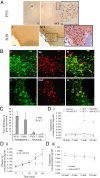
Hypothalamic neuronal activation after secretin (SCT) treatment. (A) Fos-IR in the rat paraventricular nucleus (PVN) and supraoptic nucleus (SON) 1 h after intracerebroventricular administration of vehicle (C) or SCT. Low levels of Fos-IR were observed in the PVN and SON after saline injection (C). Injection of 0.45 μg of SCT, however, induced Fos expression in magnocellular neurons of both the PVN and the SON. 3V, third ventricle. (B) Localization of SCT-induced Fos (F) in Vp- and Oxt-containing magnocellular neurons. Fos-IR was observed in the nuclei of Vp-expressing cells (arrows). M, merged image. (C) Secretin-induced changes in Vp and Oxt gene expression in the rat hypothalamic PVN and SON. Values are shown as means ± SEM fold changes compared with expression levels of Vp or Oxt in control animals (PBS-infused control group, n = 3; SCT-infused group, n = 4). (D) Effects of SCT on the release of Vp in vitro and in vivo. (Di) SCT stimulates Vp release from rat hypothalamic explants. This effect is specific to SCT and is mediated via a PKA-dependent pathway, because it was abrogated in the presence of the SCT antagonist secretin-(5–27) or the PKA inhibitor H89. After a 40-min preequilibrium period and two 5-min incubations (10 and 5 min) in normal artificial cerebrospinal fluid (ACSF) medium to determine basal release, the explants in treatment group were stimulated with 100 nM SCT (5 and 10 min). For the control, explants were incubated in ACSF solution at all time points. (Dii) Centrally injected SCT triggers Vp release into peripheral circulation by sampling blood from the right jugular vein. The Vp levels after SCT injection were compared with the baseline level at time 0, *, P < 0.01. (Diii) SCT is not able to trigger Vp release from rat pituitary explants. Experimental conditions were the same as those in Di.
Fig. 3.
Fig. 3.
Up-regulation and release of SCT in or from the hypothalamo–neurohypophysial axis during plasma hyperosmolality. (A) Up-regulation of SCT and SCTR expression in the hypothalamus and pituitary after water deprivation. Data are presented as means ± SEM. Asterisks indicate statistically significant differences (P < 0.05) when the water-deprived animals were compared with control animals. (B) Effects of chronic hyperosmolality on daily plasma SCT levels. Tap water was available ad libitum to the control group, whereas restricted water access or 0.9% saline was given to the treatment group. Blood was withdrawn daily from the tail to prepare plasma for the measurement of SCT using a rat SCT enzyme immunoassay kit (n = 6–9). *, P < 0.05 and **, P < 0.01 vs. basal value. (C) Release of SCT from rat pituitaries in response to depolarization with 80 mM K+. (Ci) Neuronal depolarization triggers SCT release from pituitary explants. After a 40-min preequilibrium period and two 5-min incubations (10 and 5 min) in normal artificial cerebrospinal fluid (ACSF) medium to determine basal release, the explants in the treatment group were stimulated for 5 min with 80 mM K+. Control experiments were performed without K+ treatment. Basal release was found to be relatively constant over time. (Cii) Action-potential- and Ca2+-mediated release of SCT from the pituitary. Outflow of SCT from pituitary explants was evoked by 80 mM K+ alone or in the presence of toxin or channel blocker. The toxins used were 1 μM TTX, 100 μM high-voltage-activated calcium channel blocker CdCl2 (Cd2+), 100 μM low-voltage-activated calcium channel blocker NiCl2 (Ni2+), 90 nM ω-agatoxin IVA (ω-Aga), 5 μM nicardipine, 300 nM Q-type calcium channel blocker ω-conotoxin MVIIC (MVIIC), 100 nM ω-conotoxin GVIA (GVIA), and 30 nM SNX-482. Results are presented in mean fold changes ± SEM of three to five determinations each in triplicate. Comparison of treated groups and controls was based on ANOVA for multiple comparisons followed by the Student–Newman–Keuls test. *, P < 0.05 and **, P < 0.01 vs. basal SCT outflow; *, P < 0.05 and **, P < 0.01 vs. K+-evoked SCT outflow. (D) Secretin levels in circulation upon stimulation of the PVN. (Di) Blood samples (150 μL) were collected for a duration of 5 min from the jugular vein through an indwelling catheter after 2 min of repeated monopolar pulse stimulation at various current intensities (0.1-ms pulses at 50 Hz, 10 s on, 10 s off, 100–500 μA). (Dii) Blood samples (150 μL) were collected every 4 min from the jugular vein from 12 min before to 44 min (4-min interval) after a 200-μA monopolar pulse stimulation (indicated with an arrow; 0.1-ms pulses at 50 Hz, 10 s) of the PVN. *, P < 0.05; **, P < 0.01.

References

    1. Jeon US, et al. Oxytocin induces apical and basolateral redistribution of aquaporin-2 in rat kidney. Nephron Exp Nephrol. 2003;93:e36–e45. - PubMed
    1. Villanger O, Veel T, Raeder MG. Secretin causes H+/HCO3− secretion from pig pancreatic ductules by vacuolar-type H+-adenosine triphosphatase. Gastroenterology. 1995;108:850–859. - PubMed
    1. Marinelli RA, Pham L, Agre P, LaRusso NF. Secretin promotes osmotic water transport in rat cholangiocytes by increasing aquaporin-1 water channels in plasma membrane. Evidence for a secretin-induced vesicular translocation of aquaporin-1. J Biol Chem. 1997;272:12984–12988. - PubMed
    1. Tietz PS, et al. Agonist-induced coordinated trafficking of functionally related transport proteins for water and ions in cholangiocytes. J Biol Chem. 2003;278:20413–20419. - PubMed
    1. Chow BK, et al. Secretin controls anion secretion in the rat epididymis in an autocrine/paracrine fashion. Biol Reprod. 2004;70:1594–1599. - PubMed

Publication types

MeSH terms