. 2009 Sep 22;106(38):16321-6.
doi: 10.1073/pnas.0906997106. Epub 2009 Sep 9.

A polycomb repressive complex 2 gene regulates apogamy and gives evolutionary insights into early land plant evolution

Affiliations

A polycomb repressive complex 2 gene regulates apogamy and gives evolutionary insights into early land plant evolution

Yosuke Okano et al. Proc Natl Acad Sci U S A. .

Abstract

Land plants have distinct developmental programs in haploid (gametophyte) and diploid (sporophyte) generations. Although usually the two programs strictly alternate at fertilization and meiosis, one program can be induced during the other program. In a process called apogamy, cells of the gametophyte other than the egg cell initiate sporophyte development. Here, we report for the moss Physcomitrella patens that apogamy resulted from deletion of the gene orthologous to the Arabidopsis thaliana CURLY LEAF (PpCLF), which encodes a component of polycomb repressive complex 2 (PRC2). In the deletion lines, a gametophytic vegetative cell frequently gave rise to a sporophyte-like body. This body grew indeterminately from an apical cell with the character of a sporophytic pluripotent stem cell but did not form a sporangium. Furthermore, with continued culture, the sporophyte-like body branched. Sporophyte branching is almost unknown among extant bryophytes. When PpCLF was expressed in the deletion lines once the sporophyte-like bodies had formed, pluripotent stem cell activity was arrested and a sporangium-like organ formed. Supported by the observed pattern of PpCLF expression, these results demonstrate that, in the gametophyte, PpCLF represses initiation of a sporophytic pluripotent stem cell and, in the sporophyte, represses that stem cell activity and induces reproductive organ development. In land plants, branching, along with indeterminate apical growth and delayed initiation of spore-bearing reproductive organs, were conspicuous innovations for the evolution of a dominant sporophyte plant body. Our study provides insights into the role of PRC2 gene regulation for sustaining evolutionary innovation in land plants.


Conflict of interest statement

The authors declare no conflict of interest.

Figures
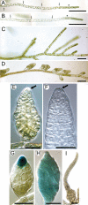
Fig. 1.
Fig. 1.
A PpCLF deletion line (ppclf-del-3) forms sporophyte-like bodies as side branches. (A and B) Wild-type (A) and ppclf-del-3 (B) protonemata grown under red light for 7 days. Asterisks and arrows indicate apical cells and septa, respectively. (C and D) Protonema of the wild type (C) and ppclf-del-3 (D) grown in white light for 2 days after 7 days of culture in red light. (E and F) Sporophyte-like body formed on the ppclf-del-3 protonema grown under white light for 7 days without (E) or with (F) clearing. Arrows indicate the apical cell. (G and H) GUS activity in sporophyte-like tissue of the ppclf/MKN4-GUS-3–1 (G) and ppclf/PpLFY2-GUS-1–1 (H) lines. (I) Sporophyte-like tissue of ppclf-del-3 grown in white light for 14 days after 7 days in red light. [Scale bars, (A–D) 100 μm; (E–I) 50 μm.]
Fig. 2.
Fig. 2.
PpCLF-Citrine is expressed in protonema and gametophore apical cells but not in sporophyte apical cells. (A and A′) Protonema apical cell. (B and B′) The third protonema cell from the apical cell. (C and C′) Side-branch initial cell. (D and D′) Gametophore stem cell. (E and E′) Gametophore tip. An arrow indicates the gametophore apical cell. (F and F′) Young antheridium. (G and G′) Mature antheridium including sperm. (H and H′) Unopened archegonium and an egg cell. (I and I′) Zygote in an opened archegonium. (J–K′) Sporophytes with an active sporophyte apical cell after the first (J and J′) and fifth (K and K′) zygotic cell division. (L–O) Sporophyte once its apical cell had stopped division and sister cells commenced division. (P–Q) Sporangium with archesporial cells. (R and R′) Spore. (S and S′) Germinating spore. (N, O, and Q) show the boxed regions in (M′ and P′). Arrows (A′, B′, C′, and H′) indicate a nucleus. For each image pair, left-hand panel is bright-field, right-hand panel is fluorescence, with the citrine signal in green and chloroplasts in magenta or white. Fluorescence of chloroplasts was detected even with the barrier filter because the fluorescence of PpCLF-Citrine was relatively weak and the exposure time needed was relatively long. [Scale bars, (A–D′, F–L′, N, O, and Q–S′) 25 μm; (E, E′, M, M′, P, and P′) 100 μm.]
Fig. 3.
Fig. 3.
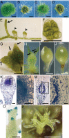
A sporangium-like organ formed following to exogenous PpCLF induction in the PpCLF deletion line. (A–D) Colonies of wild type (A), ppclf-del-3 (B), and HSP-PpCLF-Cerulean/ppclf-del-2 (C and D) grown in white light for 10 days with (A–C) or without (D) heat-shock treatment. (E) Incipient protonema (arrow) and gametophores (arrowheads) in the HSP-PpCLF-Cerulean/ppclf-del-2 line grown in white light for 2 days after 7 days in red light with heat-shock treatment. (F) A young gametophore before forming leaves in the HSP-PpCLF-Cerulean/ppclf-del-2 line, grown with heat-shock treatment for 7 days in red light and then 3 days in white light. (G and H). Sporophyte-like bodies (arrows) formed in the HSP-PpCLF-Cerulean/ppclf-del-2 line on heat-shock induced gametophores 8 days after cessation of heat-shock treatment. A young gametophore without leaves (G) and a leafy gametophore (H). (I) Sporangium-like organ of the HSP-PpCLF-Cerulean/ppclf-del-2 line grown in white light for 10 days with heat-shock treatment. (J) Wild-type sporangium. (K–N) Toluidine blue-stained sections of a sporangium-like organ of the HSP-PpCLF-Cerulean/ppclf-del-2 line (K and L) and of wild-type sporangium (M and N). (L and N) show the boxed regions in (K) and (M), respectively. (O) GUS activity in the branched sporophyte-like bodies in the ppclf/MKN4-GUS-3–1 line. (P) Sporophyte-like body with numerous branches (ppclf-del-3). [Scale bars, (A–D) 1 mm; (E, F, G, H, K, M, and O) 100 μm; (I and J) 200 μm; (L and N) 25 μm; (P) 0.5 mm.]

References

    1. Koltunow AM, Grossniklaus U. Apomixis: A developmental perspective. Annu Rev Plant Biol. 2003;54:547–574. - PubMed
    1. Bicknell RA, Koltunow AM. Understanding apomixis: Recent advances and remaining conundrums. Plant Cell. 2004;16:S228–S245. - PMC - PubMed
    1. Asker SE, Jerling L. Apomixis in Plants. Boca Raton, FL: CRC Press; 1992.
    1. Yang H-Y, Zhou C. Experimental plant reproductive biology and reproductive cell manipulation in higher plants: Now and the future. Am J Bot. 1992;79:354–363.
    1. Gifford EM, Foster AS. Morphology and Evolution of Vascular Plants. 3rd Ed. Freeman and Co.: New York; 1989.

Publication types

Associated data