. 2009 Sep 22;106(38):16351-6.
doi: 10.1073/pnas.0906922106. Epub 2009 Sep 8.

XMRV is present in malignant prostatic epithelium and is associated with prostate cancer, especially high-grade tumors

Affiliations

XMRV is present in malignant prostatic epithelium and is associated with prostate cancer, especially high-grade tumors

Robert Schlaberg et al. Proc Natl Acad Sci U S A. .

Retraction in

Abstract

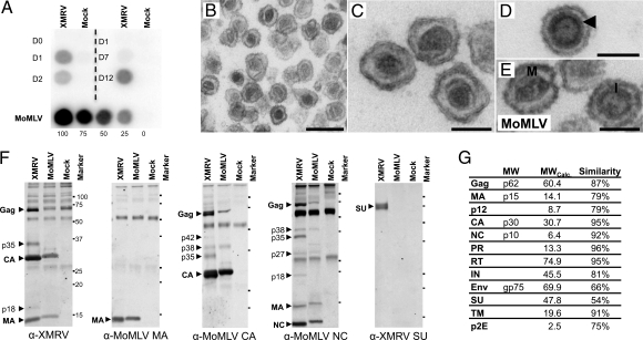
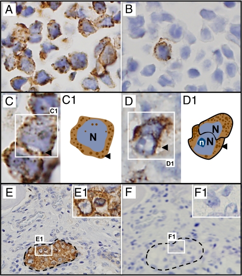
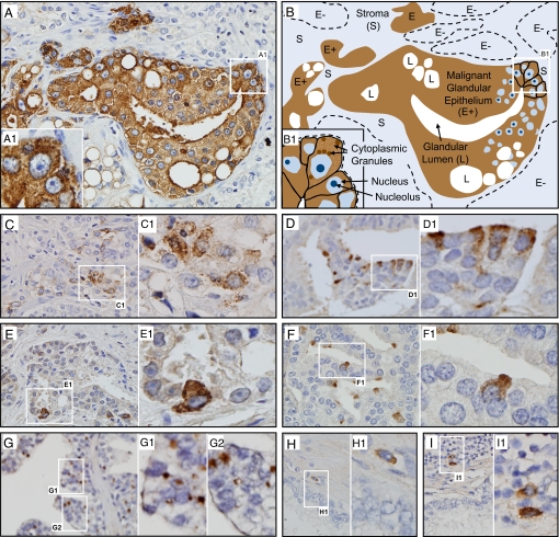
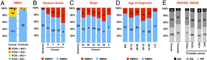
Xenotropic murine leukemia virus-related virus (XMRV) was recently discovered in human prostate cancers and is the first gammaretrovirus known to infect humans. While gammaretroviruses have well-characterized oncogenic effects in animals, they have not been shown to cause human cancers. We provide experimental evidence that XMRV is indeed a gammaretrovirus with protein composition and particle ultrastructure highly similar to Moloney murine leukemia virus (MoMLV), another gammaretrovirus. We analyzed 334 consecutive prostate resection specimens, using a quantitative PCR assay and immunohistochemistry (IHC) with an anti-XMRV specific antiserum. We found XMRV DNA in 6% and XMRV protein expression in 23% of prostate cancers. XMRV proteins were expressed primarily in malignant epithelial cells, suggesting that retroviral infection may be directly linked to tumorigenesis. XMRV infection was associated with prostate cancer, especially higher-grade cancers. We found XMRV infection to be independent of a common polymorphism in the RNASEL gene, unlike results previously reported. This finding increases the population at risk for XMRV infection from only those homozygous for the RNASEL variant to all individuals. Our observations provide evidence for an association of XMRV with malignant cells and with more aggressive tumors.


Conflict of interest statement

The authors declare no conflict of interest.

Figures

Fig. 1.
Fig. 1.
The XMRV molecular clone produces infectious particles with morphology and composition similar to MoMLV. (A) Viral release from cells transfected or inoculated with pXMRV1 or XMRV, respectively. (Left) Reverse transcriptase (RT) activity in culture supernatants from cells transfected with pXMRV1 or control EGFP plasmid. (Right) RT activity from LNCaP cells inoculated with XMRV. (Lower) RT activity from NIH 3T3 cells chronically infected with MoMLV shown for comparison. (B–E) Transmission electron microscopy of XMRV particles (B), mature XMRV cores (C), immature XMRV core, with “railroad track” marked by arrowhead (D), and MoMLV particles with mature (“M”) and immature (“I”) cores (E). (F) Western blot analysis of lysed XMRV and MoMLV virions, using antisera to XMRV whole virus, MoMLV-CA, MoMLV-MA, MoMLV-NC, and XMRV-Env SU. Comparison of blots allows identification of intermediates of Gag proteolysis, e.g., p27 (MA-p12), p42 (p12-CA), and p38 (CA-NC). (G) Molecular weights of XMRV proteins as calculated by Western blot analysis and by sequence prediction and similarity between XMRV and MoMLV proteins. [Scale bars: 250 nm (B) and 100 nm (C–E).]
Fig. 2.
Fig. 2.
XMRV proteins detected in infected cultured cells and in prostate cancer tissue by IHC, using anti-XMRV antiserum. Counterstaining with hematoxylin reveals blue nuclei. (A and B) XMRV-infected cells: 100% infected (A) and 1% infected (B). (C) Cultured infected cells at higher magnification show cytoplasmic granular staining, represented diagrammatically in C1 (arrowhead, granules). (D–F) Human prostate cancers with clusters of malignant epithelial cells (E), with Inset at higher magnification (E1). Granular staining pattern seen at higher magnification. (F and F1) Adjacent section stained with preimmune serum from the same rabbit. N, nucleus; n, nucleolus.
Fig. 3.
Fig. 3.
XMRV proteins are expressed primarily in malignant epithelial cells and very rarely in stromal cells. (A and B) IHC of a section from a qPCR-positive prostate cancer (A) and its diagrammatic representation (B). Nuclei of malignant cells are large and contain ≥1 large nucleoli (B). Multiple acini of malignant epithelial cells (E+) stain positive. All cells within these acini show intense staining. The stroma (S) and a few other acini (E−) are unstained. Insets (A1 and B1) show corresponding fields at higher magnification, with granular cytoplasmic staining pattern in several malignant epithelial cells. (C) A different field from the same sample as in A shows the range of XMRV protein expression in various acini: fewer cells expressing less protein but the same granular staining pattern. (D–F) Three additional representative samples with different frequencies of malignant epithelial cell clusters and different extents of XMRV protein expression. The intracellular staining pattern remains granular in all. (G) Staining limited to part of the cytoplasm of malignant epithelial cells in a subset of samples, as in this sample from which the XMRV clone VP62 was isolated, courtesy of R. H. Silverman and C. Magi-Galluzzi, Cleveland Clinic (6). (H and I) Scattered rare stromal cells showing cytoplasmic staining were seen close to malignant cells (H) or within inflammatory infiltrates (I).
Fig. 4.
Fig. 4.
XMRV DNA and proteins were more prevalent in prostate cancer than in controls, and especially frequent in high-grade cancers, and there was no correlation between presence of XMRV and any particular RNASEL genotype. (A) Number of prostate cancers or controls that were positive or negative for XMRV, either by qPCR or by IHC. (B–D) XMRV-positive cases (by either IHC or qPCR) correlated with Gleason grades (B), tumor stage (C), or age at diagnosis (D). (E) Presence of XMRV DNA or protein and the RNASEL genotype. Relative frequencies of RR, RQ, and QQ alleles in RNASEL at residue 462 were compared in prostate cancer cases and controls (Left), in cancers that tested positive or negative for XMRV DNA by qPCR (Center), and in cancers that tested positive or negative for XMRV proteins by IHC (Right). Cases are shown as percentages of total on the y axis and as number of cases within columns.

Comment in

References

    1. Jemal A, et al. Cancer statistics, 2008. CA Cancer J Clin. 2008;58(2):71–96. - PubMed
    1. Hayat MJ, Howlader N, Reichman ME, Edwards BK. Cancer statistics, trends, and multiple primary cancer analyses from the Surveillance, Epidemiology, and End Results (SEER) Program. Oncologist. 2007;12(1):20–37. - PubMed
    1. Parkin DM, Bray FI, Devesa SS. Cancer burden in the year 2000. The global picture. Eur J Cancer. 2001;37(Suppl 8):S4–S66. - PubMed
    1. De Marzo AM, et al. Inflammation in prostate carcinogenesis. Nat Rev Cancer. 2007;7(4):256–269. - PMC - PubMed
    1. Hayes RB, et al. Sexual behaviour, STDs and risks for prostate cancer. Br J Cancer. 2000;82(3):718–725. - PMC - PubMed

Publication types

MeSH terms