. 2009 Sep 22;106(38):16434-45.
doi: 10.1073/pnas.0905696106. Epub 2009 Sep 10.

Diminished dosage of 22q11 genes disrupts neurogenesis and cortical development in a mouse model of 22q11 deletion/DiGeorge syndrome

Affiliations

Diminished dosage of 22q11 genes disrupts neurogenesis and cortical development in a mouse model of 22q11 deletion/DiGeorge syndrome

Daniel W Meechan et al. Proc Natl Acad Sci U S A. .

Abstract

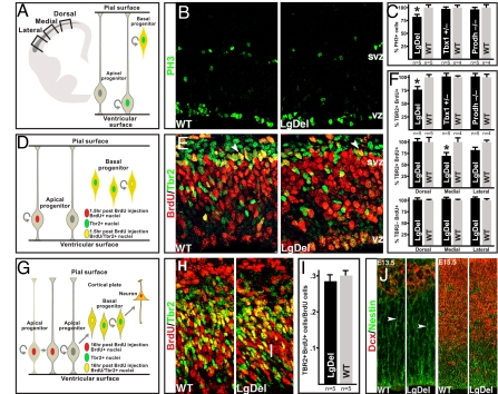
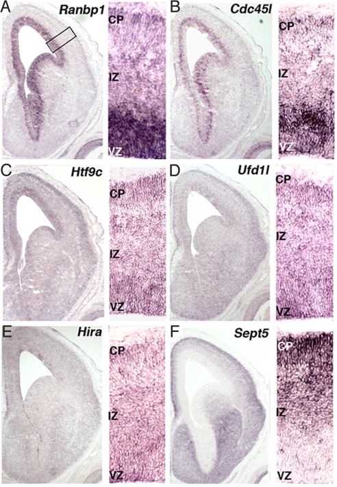
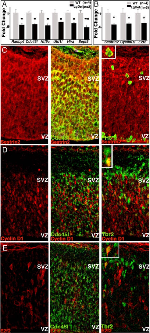
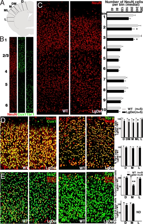
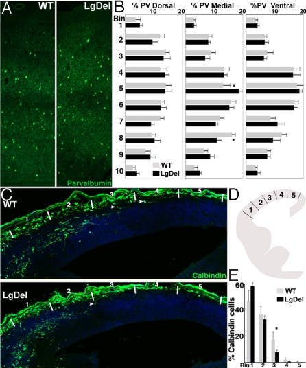
The 22q11 deletion (or DiGeorge) syndrome (22q11DS), the result of a 1.5- to 3-megabase hemizygous deletion on human chromosome 22, results in dramatically increased susceptibility for "diseases of cortical connectivity" thought to arise during development, including schizophrenia and autism. We show that diminished dosage of the genes deleted in the 1.5-megabase 22q11 minimal critical deleted region in a mouse model of 22q11DS specifically compromises neurogenesis and subsequent differentiation in the cerebral cortex. Proliferation of basal, but not apical, progenitors is disrupted, and subsequently, the frequency of layer 2/3, but not layer 5/6, projection neurons is altered. This change is paralleled by aberrant distribution of parvalbumin-labeled interneurons in upper and lower cortical layers. Deletion of Tbx1 or Prodh (22q11 genes independently associated with 22q11DS phenotypes) does not similarly disrupt basal progenitors. However, expression analysis implicates additional 22q11 genes that are selectively expressed in cortical precursors. Thus, diminished 22q11 gene dosage disrupts cortical neurogenesis and interneuron migration. Such developmental disruption may alter cortical circuitry and establish vulnerability for developmental disorders, including schizophrenia and autism.


Conflict of interest statement

The authors declare no conflict of interest.

Figures

Fig. 1.
Fig. 1.
Diminished 22q11 gene dosage disrupts cortical basal progenitor proliferation. (A) Schematic representation of a coronal section through the mouse E13.5 forebrain with the cortical SVZ indicated in gray. Proliferating cells were counted at probe locations shown by boxes, or throughout the entire cortex (Left). Schematic representation showing that basal and apical progenitors, both labeled by PH3, are discerned by their positions in the SVZ versus VZ (Right). (B) PH3 immunolabeling in the E13.5 cortex of WT and Large Deletion mice (LgDel) (10). (C) PH3 labeled cell frequency in the SVZ throughout its entire lateral to dorsal extent is significantly reduced in the LgDel cortex (*, P ≤ 0.05). However, in Prodh−/− deficient or Tbx1+/− mutants, frequency is unchanged. (D) Schematic representation of short-pulse BrdU paradigm used to label the S-phase SVZ progenitors. Dual BrdU/Tbr2 immunolabeling allows assessment of S-phase basal (Tbr2+/BrdU+) as well as apical (Tbr2-/BrdU+) progenitor frequency. (E) BrdU (90-min exposure) and Tbr2 immunolabeling in the WT and LgDel E13.5 cortex (arrowheads: double-labeled cells). (F) Tbr2+/BrdU+ SVZ cells, throughout the entire cortex, are more frequent in WT versus LgDel (*, P ≤ 0.05). However, their frequency is not changed in Prodh−/− or Tbx1−/+ (Top). There is a significant decrease of Tbr2+/BrdU+ cells in the medial and a trend in the lateral E13.5 LgDel cortex (Middle). Tbr2-/BrdU+ cell frequency is unchanged at all WT and LgDel E13.5 cortical probe locations (Lower). (G) BrdU paradigm used to assess apical progenitor production of basal progenitors. The 16-h BrdU exposure provides enough time for a fraction of labeled apical progenitor progeny to migrate toward the SVZ, and express Tbr2. Concurrently, BrdU-labeled basal progenitors down-regulate Tbr2 and migrate toward the cortical plate. The fraction of BrdU+ cells that are also Tbr2+ reflect basal progenitor generation by apical progenitors (54). (H) Tbr2+/16 h BrdU+ cells (yellow) in WT and LgDel E13.5 cortex. (I) Apical progenitor genesis of basal progenitors (Tbr2+BrdU/BrdU cells) is not significantly different between E13.5 WT and LgDel cortex (medial probe). (J) Immunofluorescent labeling in E13.5 and E15.5 WT and LgDel cortex show no disruption of migrating neuroblasts (Dcx, doublecortin) or radial glial processes (nestin) (arrowheads).
Fig. 2.
Fig. 2.
Expression localization of 22q11 cell-cycle genes during cortical neurogenesis. In all images, the entire cortical hemisphere from an E14.5 WT embryo is shown (Left), whereas higher magnification of VZ, intermediate zone (IZ), and cortical plate (CP) is shown (Right). ISH shows that 22q11 cell-cycle genes are enhanced in the VZ [Ranbp1 (A); Cdc45l (B)] enhanced in both the VZ and CP [Htf9c (C); Ufd1l (D)] lightly, but broadly expressed [Hira (E)] or enhanced in the CP [Sept5 (F)].
Fig. 3.
Fig. 3.
Changes in cell-cycle gene expression in embryonic LgDel cortex. (A) Six 22q11 putative cell-cycle genes show diminished expression by ≈50% (*, P ≤ 0.05; **, P ≤ 0.001) relative to WT E13.5 cortex. (B) Quantitative PCR verifies that three cell-cycle gene transcripts are diminished by ≈50% (*, P ≤ 0.05) in E13.5 LgDel cortex relative to WT as suggested by cell-cycle array. (C–E) Protein products of these three genes are detected in cells that also express Cdc45l, PH3, or Tbr2. (C) Sestrin2 (Left), colabeled with Cd45l (Center), or PH3 (Right). (D) CyclinD1 in E13.5 cortex (Left), colabeled with Cdc45l (Center), or Tbr2 (Right). (E) E2f2 in E13.5 cortex (Left), colabeled with Cdc45l (Center), or Tbr2 (Right).
Fig. 4.
Fig. 4.
Altered frequency of supragranular projection neurons accompanies disrupted basal progenitor proliferation. (A) Schematic representation of probe locations for quantifying frequency of (B) pan-neuronal (NeuN), layer 2–4 (Cux1), and layer 5/6 (Tbr1) cortical neurons in the WT and LgDel cortex at P5. (C) NeuN labeled cells in medial WT and LgDel probes (Left). The frequency of NeuN labeled cells across 10 equal bins from pia to white matter at the medial probe (two-way ANOVA, P ≤ 0.01, n = 5) (Right). Posthoc LSD test indicates significant reductions at LgDel bins 2 and 3 (*, P ≤ 0.05), which include supragranular cortical layers (layer 2–4). (D) Layer 2–4 neurons double-labeled for NeuN and Cux1 in P5 WT and LgDel cortex (Left). Layer 5/6 neurons double-labeled for NeuN and Tbr1 in P5 WT and LgDel cortex (Right). There is a significant decrease in Cux1 labeled neurons at dorsomedial, medial, and mediolateral cortical locations in the LgDel mutant (Upper; *, P ≤ 0.05) Tbr1 cells are not significantly altered at any cortical location (Lower). (E) Layer 2–4 neurons in P5 mouse cortex BrdU birth-dated at E13.5 (Left) and E18.5 (Right) and double-labeled for Cux1. There is a significant decrease in frequency of E13.5 generated Cux1 neurons only at the medial location in the LgDel cortex (Upper; **, P ≤ 0.01). There is no significant difference in E18.5 generated Cux1 cells (Lower; ND, not detected). D, dorsal; DM, dorsomedial; M, medial; ML, mediolateral; L, lateral.
Fig. 5.
Fig. 5.
Interneuron distribution is disrupted in the LgDel cortex. (A) Immunolabeling for parvalbumin in P21 WT and LgDel cortex (medial probe) shows altered location of cells in the LgDel cortex. (B) Parvalbumin cell distribution across 10 equivalent bins from pia to white matter in WT and LgDel P21 cortex. Cells were counted in dorsal, medial, and lateral probes. A significant interaction was observed between genotype and bin location medially (two-way ANOVA, P = 0.03, n = 5 per genotype). LSD test records a significant difference in frequency between genotypes at bins 5 and 8 (*, P ≤ 0.05). (C) Calbindin-labeled migrating interneurons in E13.5 WT and LgDel cortex. White lines mark bin locations for counting. (D) The E13.5 cortex was divided into five equidistant bins from the corticostriatal boundary to cortical hem and calbindin cell numbers in each bin determined. (E) The distribution of calbindin cells across five bins in LgDel and WT E13.5 cortex. Genotype has a significant effect on calbindin cell distribution (two-way ANOVA, *, P ≤ 0.05, n = 5 per genotype). A significant difference in calbindin frequency between genotypes at bin 3 was seen (LSD; *, P ≤ 0.05).

References

    1. Weinberger DR. Implications of normal brain development for the pathogenesis of schizophrenia. Arch Gen Psychiatry. 1987;44:660–669. - PubMed
    1. Geschwind DH, Levitt P. Autism spectrum disorders: Developmental disconnection syndromes. Curr Opin Neurobiol. 2007;17:103–111. - PubMed
    1. Bassett AS, et al. Clinical features of 78 adults with 22q11 deletion syndrome. Am J Med Genet A. 2005;138:307–313. - PMC - PubMed
    1. Murphy KC, Jones LA, Owen MJ. High rates of schizophrenia in adults with velo-cardio-facial syndrome. Arch Gen Psychiatry. 1999;56:940–945. - PubMed
    1. Niklasson L, Rasmussen P, Oskarsdottir S, Gillberg C. Autism, ADHD, mental retardation and behavior problems in 100 individuals with 22q11 deletion syndrome. Res Dev Disabil. 2009;30:763–773. - PubMed

Publication types

MeSH terms

LinkOut - more resources