. 2009 Sep 29;106(39):16657-62.
doi: 10.1073/pnas.0903775106. Epub 2009 Sep 15.

Function of IRE1 alpha in the placenta is essential for placental development and embryonic viability

Affiliations

Function of IRE1 alpha in the placenta is essential for placental development and embryonic viability

Takao Iwawaki et al. Proc Natl Acad Sci U S A. .

Abstract

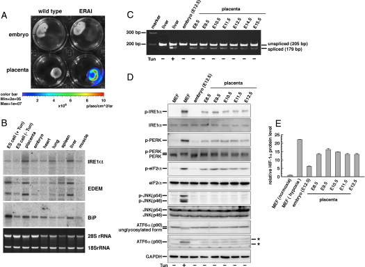
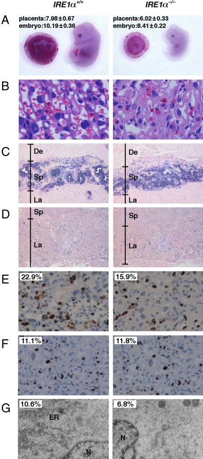
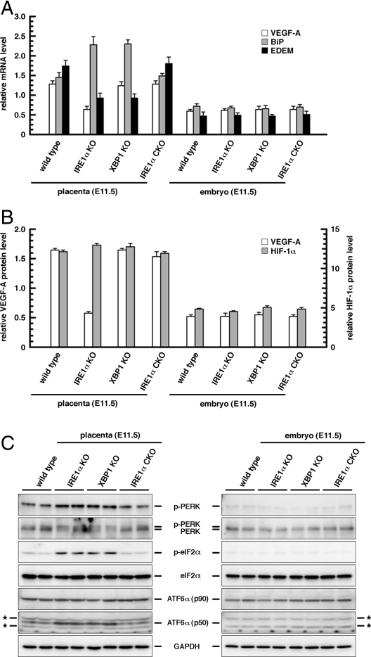
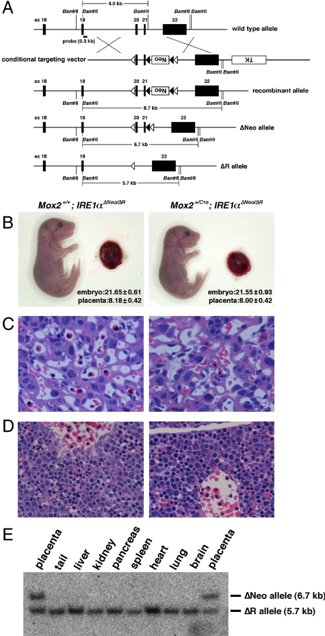
Inositol requiring enzyme-1 (IRE1), a protein located on the endoplasmic reticulum (ER) membrane, is highly conserved from yeast to humans. This protein is activated during ER stress and induces cellular adaptive responses to the stress. In mice, IRE1alpha inactivation results in widespread developmental defects, leading to embryonic death after 12.5 days of gestation. However, the cause of this embryonic lethality is not fully understood. Here, by using in vivo imaging analysis and conventional knockout mice, respectively, we showed that IRE1alpha was activated predominantly in the placenta and that loss of IRE1alpha led to reduction in vascular endothelial growth factor-A and severe dysfunction of the labyrinth in the placenta, a highly developed tissue of blood vessels. We also used a conditional knockout strategy to demonstrate that IRE1alpha-deficient embryos supplied with functionally normal placentas can be born alive. Fetal liver hypoplasia thought to be responsible for the embryonic lethality of IRE1alpha-null mice was virtually absent in rescued IRE1alpha-null pups. These findings reveal that IRE1alpha plays an essential function in extraembryonic tissues and highlight the relationship of physiological ER stress and angiogenesis in the placenta during pregnancy in mammals.


Conflict of interest statement

The authors declare no conflict of interest.

Figures

Fig. 1.
Fig. 1.
Activity and expression of IRE1α in the placenta. (A) Bioluminescence imaging of embryos and placentas of wild-type and ERAI-LUC mice (E14.5). (B) Northern blot analysis of IRE1α, EDEM, and BiP in the embryo (E11.5), placenta (E11.5), and various tissues of the adult wild-type mouse. +Tun and −Tun indicate treatment with and without 2 μg/mL tunicamycin (ER stressor) for 6 h, respectively. ES cells were used as control samples. Ethidium bromide staining (Bottom) shows the RNA loading control. (C) RT-PCR analysis of XBP1 in embryos and placentas. +Tun indicates i.p. injection with tunicamycin (500 ng/g body weight) 16 h before tissue collection. Liver tissues were used as control samples. (D) Western blot analysis of IRE1α, PERK, eIF2α, JNK, and ATF6α in embryos and placentas. p- indicates the phosphorylated form of each protein. ATF6α (p50) is the cleaved form. +Tun indicates treatment with 2.5 μg/mL tunicamycin for 6 h. MEFs were used as control samples. GAPDH was used as an internal standard. *, Nonspecific signals. (E) ELISA of HIF-1α in embryos and placentas. Normoxia and hypoxia indicate culture under 20% O2 or 1% O2 for 6 h, respectively. MEFs were used as control samples. Error bar indicates SEM (n = 3).
Fig. 2.
Fig. 2.
Placental dysplasia in IRE1α−/− mice. (A) Gross morphology of E13.5 IRE1α+/+ and IRE1α−/− embryos and placentas. Each number indicates the body length of embryo and the diameter of placenta (millimeters) as mean ± SD (n = 16). (B) H&E-stained E13.5 placental sections showing a region of the labyrinth layer in the placenta. (C and D) Cell lineage marker analysis of E13.5 placental sections by using in situ hybridization. Tpbp (C) and Tfeb (D) were used as a spongiotrophoblast cell-specific and a labyrinth cell-specific marker, respectively. De, deciduas; Sp, spongiotrophoblast; La, labyrinth. Sections were counterstained with eosin. (E) BrdU immunohistochemically stained E13.5 placental sections. Sections were counterstained with hematoxylin. Number indicates the percentage of BrdU-positive cells. (F) TUNEL assays for E13.5 placental sections. Sections were counterstained with hematoxylin. Number indicates the percentage of TUNEL-positive cells. (G) Electron micrographs of E13.5 placental sections. N, nucleus. Number indicates the percentage of the ER luminal space compared with the cytoplasmic space. Each space was measured from 10 photographs with Image Gauge version 4.21 software (Fuji film). (Original magnification: A, 1×; B, 20×; C and D, 5×; E and F, 10×; and G, 4,000×.)
Fig. 3.
Fig. 3.
Expression or activation level of VEGF-A, HIF-1α, and ER stress-responsive molecules in the placenta (E11.5) and embryo (E11.5). (A) Quantitative PCR analysis of VEGF-A, BiP, and EDEM. Error bar indicates SEM (n = 3). (B) ELISA of VEGF-A and HIF-1α. Error bar indicates SEM (n = 3). (C) Western blot analysis of PERK, eIF2α, and ATF6α. p- indicates the phosphorylated form of each protein. ATF6α (p50) is the cleaved form. GAPDH was used as an internal standard. The genotype of IRE1α CKO is Mox2+/Cre; IRE1αΔNeo/ΔR. Each sample used here was derived from the embryonic part (labyrinth and spongiotrophoblasts) of each placenta. *, Nonspecific signals.
Fig. 4.
Fig. 4.
IRE1α-deficient mice with normal placentas as reconstituted by using conditional knockout approaches. (A) Schematic diagram showing the IRE1α conditional knockout strategy. Neo and TK indicate expression units of the neomycin resistance gene and thymidine kinase gene for positive and negative selections, respectively. Closed and open arrowheads indicate FRT and loxP elements, respectively. (B) Gross morphology of E18.5 Mox2+/+; IRE1αΔNeo/ΔR and Mox2+/Cre; IRE1αΔNeo/ΔR embryos and placentas. Each number indicates the body length of embryo and the diameter of placenta (millimeters) as mean ± SD (n = 10). (C) H&E-stained E13.5 placental sections showing a region of the labyrinth layer in the placenta. (D) H&E-stained E13.5 liver sections. (Original magnifications: B, 1×; C, 20×; and D, 40×.) (E) Southern blot analysis of various tissues from Mox2+/Cre; IRE1αΔNeo/ΔR mice.

References

    1. Cox JS, Shamu CE, Walter P. Transcriptional induction of genes encoding endoplasmic reticulum resident proteins requires a transmembrane protein kinase. Cell. 1993;73:1197–1206. - PubMed
    1. Mori K, Ma W, Gething MJ, Sambrook J. A transmembrane protein with a cdc2+/CDC28-related kinase activity is required for signaling from the ER to the nucleus. Cell. 1993;74:743–756. - PubMed
    1. Cox JS, Walter P. A novel mechanism for regulating activity of a transcription factor that controls the unfolded protein response. Cell. 1996;87:391–404. - PubMed
    1. Shamu ES, Walter P. Oligomerization and phosphorylation of the Ire1p kinase during intracellular signaling from the endoplasmic reticulum to the nucleus. EMBO J. 1996;15:3028–3039. - PMC - PubMed
    1. Calfon M, et al. IRE1 couples endoplasmic reticulum load to secretory capacity by processing the XBP-1 mRNA. Nature. 2002;415:92–96. - PubMed

Publication types