. 2009 Sep 29;106(39):16669-74.
doi: 10.1073/pnas.0907138106. Epub 2009 Sep 15.

Dual role of K ATP channel C-terminal motif in membrane targeting and metabolic regulation

Affiliations

Dual role of K ATP channel C-terminal motif in membrane targeting and metabolic regulation

Crystal F Kline et al. Proc Natl Acad Sci U S A. .

Abstract

The coordinated sorting of ion channels to specific plasma membrane domains is necessary for excitable cell physiology. K(ATP) channels, assembled from pore-forming (Kir6.x) and regulatory sulfonylurea receptor subunits, are critical electrical transducers of the metabolic state of excitable tissues, including skeletal and smooth muscle, heart, brain, kidney, and pancreas. Here we show that the C-terminal domain of Kir6.2 contains a motif conferring membrane targeting in primary excitable cells. Kir6.2 lacking this motif displays aberrant channel targeting due to loss of association with the membrane adapter ankyrin-B (AnkB). Moreover, we demonstrate that this Kir6.2 C-terminal AnkB-binding motif (ABM) serves a dual role in K(ATP) channel trafficking and membrane metabolic regulation and dysfunction in these pathways results in human excitable cell disease. Thus, the K(ATP) channel ABM serves as a previously unrecognized bifunctional touch-point for grading K(ATP) channel gating and membrane targeting and may play a fundamental role in controlling excitable cell metabolic regulation.


Figures

Fig. 1.
Fig. 1.
Kir6.2 C-terminal motif binds AnkB. (A) Domain organization of KATP channel subunits Kir6.2 and SUR. (B) Kir6.2 C terminus contains a putative ankyrin-binding motif similar to motif in voltage-gated Nav channels. (C) Kir6.2 motif is highly conserved across vertebrates. (D) Kir6.2 associates with AnkB in vivo (pancreas) by co-immunoprecipitation. (E) Radiolabeled Kir6.2 directly associates with purified AnkB membrane-binding domain, but not other AnkB domains. Inset shows purified AnkB domains. (F) Kir6.2 C-terminal domain containing putative AnkB-binding motif is required for AnkB-binding. (G) Kir6.2 C-terminal motif is required for AnkB-binding. Note absence of AnkB-binding for Kir6.2 mutant Δ315 vs. Δ331. (H) Kir6.2 C-terminal residues 316–323 are sufficient for AnkB-binding activity (radiolabeled AnkB MBD). In contrast, the corresponding motif in the Kir6.1 C terminus lacks AnkB-binding activity.
Fig. 2.
Fig. 2.
Kir6.2 membrane expression requires AnkB. (A and B) AnkB is highly expressed in β cells and this expression is reduced in AnkB+/− islets. (C–J) In contrast to other β cell membrane and cytoskeletal proteins, Kir6.2 expression is reduced in cells lacking AnkB by immunoblot and immunostaining. (Scale bar, 10 μm.) (K–O) Generation and characterization of primary AnkB−/− fibroblasts. Expanded primary cultures were analyzed for ankyrin expression by PCR genotyping (K), immunoblot (L), and immunostaining (N and O; AnkB, green; phalloidin, red; nuclear dye topro-3, blue). (M) Primary fibroblasts lack endogenous Kir6.2 expression. (P–R) Kir6.2-GFP targeting requires AnkB. (P) GFP expression in WT fibroblasts. (Q) GFP-Kir6.2 expression in WT fibroblasts. (R) GFP-expression in AnkB−/− fibroblasts. (S) Expression of exogenous AnkB rescues GFP-Kir6.2 localization in AnkB−/− fibroblasts.
Fig. 3.
Fig. 3.
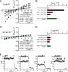
AnkB is required for Kir6.2 membrane expression. Current-voltage relationship for IKATP recorded from WT, AnkB+/−, AnkB−/− fibroblasts transfected with Kir6.2 and SUR1 using the inside-out patch clamp configuration at zero ATP (A and B) to stimulate the opening of all membrane KATP channels (n > 5, P < 0.05), and at 1 mM ATP to block IKATP (C and D). All pipets displayed similar resistances. (B and D) Summary data for IKATP current at −60 mV (B; 0 ATP; D; 1 mM ATP). Note that expression of AnkB cDNA rescues abnormal channel targeting phenotype in AnkB−/− cells. (E) IKATP recordings for WT, AnkB+/−, AnkB−/− fibroblasts, and AnkB−/− fibroblasts +AnkB cDNA recorded at −60 mV. Arrow denotes change in solution from 0 ATP to 1 mM ATP (channel closure). Note that primary cells displayed a small, endogenous K+ current (present in nontransfected cells) that was not blocked by 1 mM ATP (ATP insensitive, IKATP-independent).
Fig. 4.
Fig. 4.
Kir6.2 C-terminal acidic motif mediates channel targeting and human diabetes mutation in motif abolishes Kir6.2 trafficking. (A) Identification of human permanent neonatal diabetes mutation in Kir6.2 C-terminal motif (E322K) and alignment with Nav1.5 human Brugada syndrome arrhythmia mutation (affects ankyrin-binding and channel targeting). (B) Human PNDM mutation in Kir6.2 C-terminal motif blocks AnkB-binding. (C and D) Human PNDM mutation E322K is sufficient to reduce AnkB-binding in context of peptide. (E and F) In contrast to GFP, GFP-Kir6.2 is expressed primarily at the membrane of HEK293 cells. (G–K) GFP-Kir6.2 C-terminal motif mutants that affect AnkB-binding display abnormal membrane targeting in HEK293 cells (G–I) and primary WT (J) and AnkB−/− fibroblasts (K). Note perinuclear expression of mutants (white arrows). Additional staining represents phalloidin (red) and nuclear dye topro-3 (blue). (Scale bar, 10 μm.)
Fig. 5.
Fig. 5.
Kir6.2 C-terminal motif plays role in KATP channel metabolic regulation. (A–C) Membrane-associated KATP channels composed of E322K Kir6.2 subunits (lack ankyrin-binding) demonstrate a significant reduction in ATP sensitivity. Mutant channels demonstrate a rightward shift in ATP sensitivity (WT Kir6.2 = 10 μM, E322K channels = 62 μM) versus WT channels. (C) We observed no difference in the ATP sensitivity of Kir6.2 E322A (displays normal ankyrin-binding activity) compared with WT channels.

References

    1. Seino S. ATP-sensitive potassium channels: A model of heteromultimeric potassium channel/receptor assemblies. Annu Rev Physiol. 1999;61:337–362. - PubMed
    1. Nichols CG. KATP channels as molecular sensors of cellular metabolism. Nature. 2006;440:470–476. - PubMed
    1. Gloyn AL, Siddiqui J, Ellard S. Mutations in the genes encoding the pancreatic beta-cell KATP channel subunits Kir6.2 (KCNJ11) and SUR1 (ABCC8) in diabetes mellitus and hyperinsulinism. Hum Mutat. 2006;27:220–231. - PubMed
    1. Bahi-Buisson N, et al. Infantile spasms as an epileptic feature of DEND syndrome associated with an activating mutation in the potassium adenosine triphosphate (ATP) channel, Kir6.2. J Child Neurol. 2007;22:1147–1150. - PubMed
    1. Bienengraeber M, et al. ABCC9 mutations identified in human dilated cardiomyopathy disrupt catalytic KATP channel gating. Nat Genet. 2004;36:382–387. - PMC - PubMed

Publication types