. 2009 Dec;29(24):6438-48.
doi: 10.1128/MCB.00398-09. Epub 2009 Oct 5.

Alternative splicing modulates autoinhibition and SH3 accessibility in the Src kinase Fyn

Affiliations

Alternative splicing modulates autoinhibition and SH3 accessibility in the Src kinase Fyn

C Brignatz et al. Mol Cell Biol. 2009 Dec.

Abstract

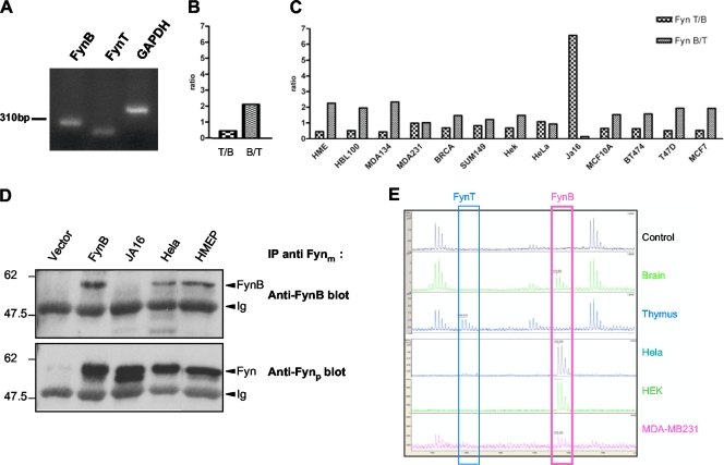
Src family kinases are central regulators of a large number of signaling pathways. To adapt to the idiosyncrasies of different cell types, these kinases may need a fine-tuning of their intrinsic molecular control mechanisms. Here, we describe on a molecular level how the Fyn kinase uses alternative splicing to adapt to different cellular environments. Using structural analysis, site-directed mutagenesis, and functional analysis, we show how the inclusion of either exon 7A or 7B affects the autoinhibition of Fyn and how this changes the SH3-dependent interaction and tyrosine phosphorylation of Sam68, with functional consequences for the Sam68-regulated survival of epithelial cells. Our results illustrate a novel mechanism of evolution that may contribute to the complexity of Src kinase regulation.


Figures

FIG. 1.
FIG. 1.
Fyn expression in epithelial cells. (A) Analysis of FynB, FynT, and GAPDH mRNAs in HMEP cells by quantitative RT-PCR. PCR products obtained after 40 cycles were analyzed on a 2% agarose gel followed by ethidium bromide staining and photography. (B) The CT values from samples obtained from experiments depicted in panel A were determined, and after normalization to results for GAPDH mRNA, FynB/FynT (B/T) and FynT/FynB (T/B) ratios were calculated and plotted. (C) The FynB/FynT and FynT/FynB mRNA ratios were determined as described for panel B in the indicated epithelial cancer cell lines (HME, HBL100, MDA-MB134, MDA-MB231, BRCA, SUM149, HEK293, HeLa, MCF10A, BT474, T47D, and MCF7) as well as in a leukemia cell line (JA16). (D) Using polyclonal anti-Fyn rabbit antibodies, the Fyn protein was immunoprecipitated (IP) from HEK293 cells transfected with empty vector or with FynB-encoding plasmid constructs or from HMEP, HeLa, and JA16 cell lines, followed by SDS-PAGE and anti-FynB and anti-Fyn immunoblotting, as indicated. Ig, immunoglobulin. (E) Fyn protein was immunoprecipitated from the indicated tissues and cell lines using polyclonal anti-Fyn rabbit antibodies, separated on SDS-PAGE, and characterized by MALDI-TOF mass spectrometry after tryptic digestion. The specific p1948 FynT (218A-233K) and p1976 FynB (218A-233R) peptides differing only by the last amino acid are framed blue and pink, respectively.
FIG. 2.
FIG. 2.
FynB- and FynT-induced tyrosine phosphorylation pattern. (A) Schematic diagram of the Fyn constructs. The red star indicates the Y528F mutation, the black star indicates the P251A mutation in SH2-kinase linker, and the FynΔSH3 construct was obtained by deleting the SH3 domain sequences. (B) HEK293 cells were transfected with plasmid constructs encoding FynB, FynB_Y528F, FynT, FynT_Y528F, FynBΔSH3, FynTΔSH3, FynB-P251A, or FynT-P251A. After 48 h of expression, cell extracts were prepared and analyzed by Western blotting for tyrosine phosphorylation (pY blot), Sam68 (Sam68 blot), and tubulin (tubulin blot) expression. (C) The extracts used for panel B were immunoprecipitated (IP) using Fyn antibodies, followed by Sam68 and Fyn immunoblotting. (D) HEK293 cells were transfected with increasing amounts of GFP-Sam68 or empty (−) plasmid constructs (0, 0.2, 0.6, 1.8, or 2.6 ng) and cotransfected with FynB or FynT (0.2 ng), as indicated. Whole-cell lysates were analyzed by Western blotting as described for panel B. (E) HEK293 cells were transfected with increasing amounts of FynB, FynT, or empty (−) plasmid constructs (0, 0.2, 0.6, 1.8, or 2.6 ng) and cotransfected with GFP-Sam68 (0.2 ng). Whole-cell lysates were analyzed by Western blotting as described for panel B. Band intensities were quantified using Image J, and the phospho-Sam68/Sam68 ratios are presented.
FIG. 3.
FIG. 3.
Specificity of the differential FynB and FynT phosphorylation activities. (A) HEK293 cells were transfected with plasmid constructs encoding Abl, c-Kit, FynT, Tec, and Ret tyrosine kinases. After 48 h of expression, cell extracts were analyzed by Western blotting for tyrosine phosphorylation (pY blot) and for the expression of each corresponding kinase, as indicated. (B) HEK293 cells were transfected with plasmid constructs encoding FynB, FynT, Src, Lck, Yes, and Hck Src kinases. After 48 h of expression, cells extracts were analyzed by Western blotting for tyrosine phosphorylation (pY blot) and Sam68 expression (Sam68 blot). (C) HEK293 cells were transfected with plasmid constructs encoding FynB and FynT. After 48 h of expression, cells extracts were analyzed by anti-phospho-Shc (TYR239/240), anti-Fyn, and anti-Shc Western blotting, as indicated. (D) HEK293 cells were transfected with plasmid constructs encoding FynB and FynT, followed by phospholipase C-γ1 immunoprecipitation (IP) and antiphosphotyrosine (pY blot), anti-Fyn, and anti-phospholipase C-γ1 Western blotting, as indicated. Total cell extracts also were analyzed for Fyn expression (lower panel). (E) HEK293 cells were transfected with plasmid constructs encoding FynB and FynT. Cell extracts were immunoprecipitated using Fyn antibody, followed by antiphosphotyrosine (pY blot) and anti-Fyn Western blotting. An asterisk indicate proteins that were phosphorylated on tyrosine residues in both FynB- and FynT-transfected cells; a plus sign indicate proteins that were phosphorylated on tyrosine residues in FynT-transfected cells but not in FynB-transfected cells.
FIG. 4.
FIG. 4.
Evidence for FynB autoinhibition that is distinct from FynT autoinhibition. (A) COS7 cells were transfected with plasmid constructs encoding FynB or FynT. Cell extracts were immunoprecipitated using Fyn rabbit polyclonal antibodies, followed by immunoblotting using specific anti-Src-pTyr416 antibody (upper panel), and anti-Src-pTyr527 (lower panel) antibodies. The position of phosphorylated-Fyn (P-Fyn) and immunoglobulin heavy chains (Ig) are indicated. WCL, whole-cell lysate. (B) Initial homology models of FynT and FynB were created using the Swiss-Model server and the c-Src structure (PDB entry 1FMK [47]) as a template. Models were manually adjusted and subjected to structure idealization. Final models show verify 3D scores that are similar to those of the experimental Src structure. The SH2-kinase linker is shown in cyan for FynT (left panel) and in blue for FynB (right panel). The SH3 domain is shown in green on the left side of the SH2-kinase linker, and SH1 domain is shown in magenta on the right side. (C) The fyn gene exon 7 sequences from different species coding for FynB and FynT proteins were aligned using ClustalW (Blosum 30 matrix). The alignment shows that the FynB linker contains more charged residues (red) and hydrophobic residues (green) than the FynT linker. The SH2 domain limit is known with the crystal of the SH2 domain of Fyn (2, 26). The SH1 domain limit is fixed based on the known Src structure (8).
FIG. 5.
FIG. 5.
Role of the SH2-SH1 linker on FynB and FynT kinase activities. (A) HEK293 cells were transfected with plasmid constructs encoding FynB, FynT, FynB-Y528F, FynT-Y528F, FynB-P251A, FynT-P251A, FynB-L256V, FynT-L256V, FynB-P251A-L256V, and FynT-P251A-L256V. Total cell extracts were analyzed by anti-phospho-SrcTyr416, anti-phospho-SrcTyr527, and anti-Fyn Western blotting, as indicated. (B) HEK293 cells were transfected with plasmid constructs encoding FynB or FynT. After 48 h of expression, cells were lysed in RIPA buffer and cell extracts were immunoprecipitated using Fyn rabbit polyclonal antibodies. Immunoprecipitates (IP) were incubated with 0, 2, or 30 μM of the p85-derived proline-rich peptide for 15 min at 30°C, followed by an in vitro kinase assay in kinase assay buffer containing enolase and 2 μCi [γ-32P]ATP for 15 min at 30°C. The reaction was terminated by adding SDS-PAGE loading buffer. Samples were analyzed by SDS-PAGE followed by autoradiography (upper panel) and Fyn immunoblotting (middle panel). The positions of phosphorylated Fyn (p-Fyn) and the phosphorylated enolase (p-Enolase) are indicated by arrows. Densitometric analyses are shown in the lower panel and were obtained with Image J software. Phosphorylated enolase signals were normalized to total Fyn levels. Results are presented as percent increases of normalized phosphorylated enolase levels.
FIG. 6.
FIG. 6.
Core SH2-kinase linker residues are both required and sufficient to confer FynB Sam68 phosphorylation that is different from that of FynT. (A) The exon 7 sequences are shown in pink for FynB and in blue for FynT. Conserved residues are shown in black. The FynB-like construct was obtained by swapping the FynB SH2-kinase linker core residues as defined in the legend to Fig. 4C (pink boxed residues) in the FynT backbone (represented by a blue line). The FynT-like construct was obtained by swapping the FynT SH2-kinase linker core residues as defined in the legend to Fig. 4C (blue boxed residues) in the FynB backbone (represented by a pink line). (B) HEK293 cells were transfected with plasmid constructs encoding FynB, FynT, FynT-like, or FynB-like. After 48 h of expression, cell extracts were analyzed by Western blotting for tyrosine phosphorylation (pY blot) and for Sam68 (Sam68 blot), Fyn (Fyn blot), and tubulin (tubulin blot) expression. (C) The extracts used for panel B were immunoprecipitated using Fyn antibodies, followed by Sam68 and Fyn immunoblotting. (D) Splicing assay of the Bcl-x minigene (0.5 μg) in HEK293 cells transfected with 0.25 μg of either GFP or GFP-Sam68 alone or in combination with 0.25 μg of FynT or FynB plasmid constructs. Cells were harvested 20 h after transfection and processed for RT-PCR (upper panel). Densitometric analyses are shown in the lower panel as mean values of the Bcl-xs/Bcl-xL ratio.
FIG. 7.
FIG. 7.
Differential regulation of Sam68-dependent Bcl-x mRNA splicing by FynB and FynT correlates with differential induction of apoptosis. (A) HEK293 cells were transfected with GFP or GFP-Sam68 and cotransfected with FynB or FynT plasmid construct (1:1 ratio) using Lipofectamine reagent. Cells were fixed and permeabilized 30 h after transfection, followed by immunofluorescence using anti-activated (cleaved) caspase 3. Cells positive for active caspase 3 among the GFP-positive cells were counted. Results are the means ± standard deviations from three independent experiments. ctrl, control.
FIG. 8.
FIG. 8.
Distinct SH3 domain ligand interaction and phosphorylation by FynB and FynT. FynB is shown in pink and FynT in blue. The tables summarize the differential FynB and FynT conformation and activation by SH3 ligands (green).

References

    1. Arnold, K., L. Bordoli, J. Kopp, and T. Schwede. 2006. The SWISS-MODEL workspace: a web-based environment for protein structure homology modelling. Bioinformatics 22:195-201. - PubMed
    1. Arold, S. T., T. S. Ulmer, T. D. Mulhern, J. M. Werner, J. E. Ladbury, I. D. Campbell, and M. E. Noble. 2001. The role of the Src homology 3-Src homology 2 interface in the regulation of Src kinases. J. Biol. Chem. 276:17199-17205. - PubMed
    1. Briggs, S. D., M. Sharkey, M. Stevenson, and T. E. Smithgall. 1997. SH3-mediated Hck tyrosine kinase activation and fibroblast transformation by the Nef protein of HIV-1. J. Biol. Chem. 272:17899-17902. - PubMed
    1. Brown, M. T., and J. A. Cooper. 1996. Regulation, substrates and functions of src. Biochim. Biophys. Acta 1287:121-149. - PubMed
    1. Busà, R., M. P. Paronetto, D. Farini, E. Pierantozzi, F. Botti, D. F. Angelini, F. Attisani, G. Vespasiani, and C. Sette. 2007. The RNA-binding protein Sam68 contributes to proliferation and survival of human prostate cancer cells. Oncogene 26:4372-4382. - PubMed

Publication types

MeSH terms

LinkOut - more resources