. 2009 Dec;29(24):6366-79.
doi: 10.1128/MCB.01259-09. Epub 2009 Oct 5.

Transcription factor Glis3, a novel critical player in the regulation of pancreatic beta-cell development and insulin gene expression

Affiliations

Transcription factor Glis3, a novel critical player in the regulation of pancreatic beta-cell development and insulin gene expression

Hong Soon Kang et al. Mol Cell Biol. 2009 Dec.

Erratum in

  • Mol Cell Biol. 2010 Apr;30(7):1864. Pierreux, Christophe E [added]; Lemaigre, Frederic P [added]

Abstract

In this study, we report that the Krüppel-like zinc finger transcription factor Gli-similar 3 (Glis3) is induced during the secondary transition of pancreatic development, a stage of cell lineage specification and extensive patterning, and that Glis3(zf/zf) mutant mice develop neonatal diabetes, evidenced by hyperglycemia and hypoinsulinemia. The Glis3(zf/zf) mutant mouse pancreas shows a dramatic loss of beta and delta cells, contrasting a smaller relative loss of alpha, PP, and epsilon cells. In addition, Glis3(zf/zf) mutant mice develop ductal cysts, while no significant changes were observed in acini. Gene expression profiling and immunofluorescent staining demonstrated that the expression of pancreatic hormones and several transcription factors important in endocrine cell development, including Ngn3, MafA, and Pdx1, were significantly decreased in the developing pancreata of Glis3(zf/zf) mutant mice. The population of pancreatic progenitors appears not to be greatly affected in Glis3(zf/zf) mutant mice; however, the number of neurogenin 3 (Ngn3)-positive endocrine cell progenitors is significantly reduced. Our study indicates that Glis3 plays a key role in cell lineage specification, particularly in the development of mature pancreatic beta cells. In addition, we provide evidence that Glis3 regulates insulin gene expression through two Glis-binding sites in its proximal promoter, indicating that Glis3 also regulates beta-cell function.


Figures

FIG. 1.
FIG. 1.
Expression of Glis3 during pancreatic development. (A) The expression of Glis3 mRNA (n = 4) during pancreatic development was examined by QRT-PCR analysis. The expression of Glis3 was normalized to glyceraldehyde-3-phosphate dehydrogenase. (B) Comparison of the relative levels of Glis3 mRNA expression in αTC (pancreatic α-cell-like line), βTC (β-cell-like line), and PANC1 (pancreatic epithelial cell-like line) cell lines and pancreatic islets. The relative expression of Glis3 was analyzed from the Genespeed database (http://genespeed.ccf.org/). (C to G) The expression of Glis3 mRNA in E11.5, E12.5, E13.5, E16.5, and E18.5 pancreata was examined by in situ hybridization. Sto, stomach; vp, ventral pancreas; dp, dorsal pancreas; Int, intestine.
FIG. 2.
FIG. 2.
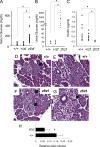
Development of hyperglycemia and hypoinsulinemia in Glis3zf/zf mutant mice. (A) Elevated blood glucose levels in WT (n = 11), Glis3+/zf mutant (n = 12), and Glis3zf/zf mutant (n = 16) mice. (B) Analysis of urine glucose levels in WT (n = 11), Glis3+/zf mutant (n = 9), and Glis3zf/zf mutant (n = 11) mice. (C) Comparison of blood insulin levels in WT (n = 10), Glis3+/zf mutant (n = 11), and Glis3zf/zf mutant (n = 14) mice. (D to G) The size of pancreatic islets is significantly reduced in Glis3zf/zf mutant mice. Shown are hematoxylin-and-eosin-stained sections of pancreata from PND3 WT and Glis3zf/zf mutant mice. Arrows indicate pancreatic islets. (H) Islet volume was reduced in the PND3 Glis3zf/zf mutant pancreas (n = 6) compared to that in the WT pancreas (n = 6). By a systematic random sampling approach, the islet load was assessed in every 10th section throughout the pancreas. An asterisk indicates a P value of <0.02.
FIG. 3.
FIG. 3.
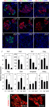
Loss of Glis3 function causes a dramatic reduction in endocrine cells, particularly β cells. (A to I) Sections of PND3 WT and Glis3+/zf and Glis3zf/zf mutant pancreata were examined by immunofluorescence with antibodies against insulin (Ins), glucagon (Gcg), pancreatic polypeptide (Ppy), and somatostatin (Sst) as indicated. Nuclei were visualized by 4′,6-diamidino-2-phenylindole (DAPI) staining. (J) The expression of several endocrine cell-specific, but not exocrine cell-specific, genes is greatly reduced in Glis3zf/zf mutant mice. RNAs isolated from pancreata of PND3 WT and Glis3+/zf and Glis3zf/zf mutant mice were analyzed by QRT-PCR for the expression of insulin 1 and 2 (Ins1 and Ins2), somatostatin, glucagon, ghrelin (Ghrl), and pancreatic polypeptide mRNAs and the exocrine cell-related genes for amylase and Ptf1a. An asterisk indicates a P value of <0.02. (K, L) Expression of amylase is not significantly altered in the Glis3zf/zf mutant mouse pancreas. Sections of E16.5 WT and Glis3zf/zf mutant mouse pancreata were stained with antibodies against the exocrine enzyme amylase (red) and the ductal marker osteopontin (Opn; green).
FIG. 4.
FIG. 4.
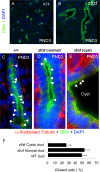
Development of dilated and cystic pancreatic ducts in Glis3zf/zf mutant mice. (A, B) Pancreata from PND3 WT and Glis3zf/zf mutant mice were stained with the ductal marker DBA and DAPI. (C to E) Pancreatic ducts and cilia were stained by DBA and anti-α-acetylated tubulin, respectively. Representative images of a pancreatic duct from a PND3 WT mouse (C) and a normal (D) and a cystic (E) pancreatic duct from a PND3 Glis3zf/zf mutant mouse are shown. In WT mice, about 50% of the ductal cells were stained positively with anti-α-acetylated tubulin antibody. The number of ciliated cells was greatly reduced in cysts of the Glis3zf/zf mutant pancreas. Arrowheads indicate primary cilia. (F) The percentage of ciliated cells is reduced in cystic pancreatic ducts of Glis3zf/zf mutant mice. Sections from three different mice in each group were selected randomly and stained with DBA and anti-α-acetylated tubulin antibody. The percentage of DBA-positive cells (n = 50 to 70) containing a primary cilium was calculated.
FIG. 5.
FIG. 5.
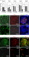
The expression of several endocrine cell-related transcription factors is significantly reduced in the Glis3zf/zf mutant pancreas. (A) The expression of Nkx6.1, Pdx1, Pax4, Ngn3, and MafA, genes associated with endocrine development, was examined by QRT-PCR in pancreas samples from PND3 WT and Glis3+/zf and Glis3zf/zf mutant mice (n = 3). An asterisk indicates a P value of <0.02. (B to M) Pancreas sections from PND3 WT and Glis3zf/zf mutant mice were examined by immunofluorescence with antibodies for Nkx6.1, Pdx1, insulin, MafA, and/or Glut2, as indicated. D, G, J, and M, merged. DAPI, blue staining.
FIG. 6.
FIG. 6.
Analysis of the expression of Pdx1, Ngn3, and glucagon at E11.5 of pancreatic development. (A to H) Expression of Ngn3, Pdx1, and glucagon in pancreas sections from E11.5 Glis3zf/zf mutant and WT embryos was examined by immunofluorescent staining. Representative images are shown. No significant differences were observed. Ngn3, green in panels A and E; Pdx1, red in panels B and F; merged in panels C and G. (D, H) Merged glucagon (Gcg; green) and Pdx1 (red) staining of E11.5 pancreas.
FIG. 7.
FIG. 7.
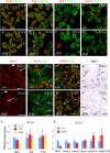
Comparison of Sox9, Pdx1, Ngn3, and HNF6 expression during the development of WT and Glis3zf/zf mutant pancreata. (A to H) The Sox9, Pdx1, glucagon, Ngn3, and E-cadherin proteins were analyzed by immunofluorescence in sections of E13.5 WT and Glis3zf/zf mutant pancreata. (I to N) The Pdx1, Ins, and Hnf6 proteins were analyzed in sections of E16.5 WT and Glis3zf/zf mutant pancreata by immunofluorescence staining as indicated. (I, J) The numbers of cells expressing high levels of Pdx1 were dramatically decreased in the Glis3zf/zf mutant pancreas, as indicated by arrows. Arrowheads indicate cells expressing low levels of Pdx1. (K, L). The number of cells expressing insulin was dramatically decreased in the Glis3zf/zf mutant pancreas, as indicated by arrows. (M, N) The expression of Hnf6 is high in the forming ducts (indicated by arrows) and low in the exocrine tissue (indicated by arrowheads); the asterisks indicate nonspecific fluorescence in vessels. (O, P) Ngn3 mRNA expression was examined by in situ hybridization in sections of E16.5 WT and Glis3zf/zf mutant pancreata. (Q) The expression of Ngn3, Gcg, and Pdx1 mRNAs was analyzed in samples from WT and Glis3+/zf and Glis3zf/zf mutant E13.5 embryos (n = 3) by QRT-PCR. (R) Expression of Ngn3, Notch1 to -3, Hnf6, and HNF6β mRNAs was compared between E15.5 WT (n = 3) and Glis3zf/zf mutant (n = 3) pancreata. Expression was analyzed by QRT-PCR. An asterisk indicates a P value of <0.05.
FIG. 8.
FIG. 8.
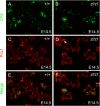
The expression of Cpa1 was not significantly changed in pancreata of WT and Glis3zf/zf mutant E14.5 embryos. Cpa1, green in panels A and B; Pdx1, red in panels C and D; merged in panels E and F.
FIG. 9.
FIG. 9.
Glis3 functions as a transcriptional regulator of insulin gene expression. (A) Schematic representation of potential Glis-BS and other enhancers in the murine and rat Ins2 and human INS 5′ regulatory regions. The scale at the top is in base pairs relative to the transcriptional start site (represented as +1). (B) Comparison of the nucleotide sequences of the consensus Glis-BS with the two putative Glis-BS in the mouse Ins2 and human INS proximal promoter regions. (C) Two putative Glis-BS-containing regions in the mouse insulin 2 promoter (Glis-BS1 at −84 to −109 and Glis-BS2 at −253 to −276) and their respective mutant nucleotides were labeled and used as probes in an EMSA. Competition was carried out with 5-fold (+) and 25-fold (++) excesses of unlabeled oligonucleotides as indicated. Arrow indicates the Glis3-Glis-BS complex. (D, E) Binding of Glis3 to 32P-labeled consensus Glis-BS was examined in the absence or presence of unlabeled consensus Glis-BS, mouse Glis-BS1, and mGlis-BS2 sites, their mutants (mut), or the corresponding human INS Glis-BS. (F) INS-1 (832/13) cells were cotransfected with the pGL4.10 empty vector, mIns2-696-Luc, or hINS-700-Luc and either the p3xFlag-CMV-10 empty vector, p3xFlag-CMV-Glis3, or p3xFlag-CMV-Glis3ΔC748 as indicated. After 24 h, cells were assayed for luciferase and β-galactosidase activities and the relative Luc activity was calculated and plotted. Each bar represents the mean ± the standard error of the mean. (G) INS-1 (832/13) cells were cotransfected with the pGL4.10 empty vector, mIns2(−696)-Luc, or the specified mIns2(−696)-Luc mutant and either p3xFlag-CMV-10, p3xFlag-CMV-Glis3, or p3xFlag-CMV-Glis3ΔN302 as indicated. Relative Luc activity was calculated and plotted. In the insert, shaded boxes indicate Glis-BS and X indicates mutated Glis-BS. (H) INS-1 (832/13) cells were transiently transfected with p3xFlag-CMV-Glis3 or p3xFlag-CMV-Glis3ΔN302 and 48 h later analyzed by QRT-PCR for the expression of rat Ins1 and Ins2 and mouse Glis3.
FIG. 10.
FIG. 10.
Schematic representation of the critical roles of Glis3 in pancreatic development and insulin gene expression. Pdx1+ Ptf1a+ Cpa1+ progenitor cells can differentiate into proendocrine, ductal, and exocrine lineages. Glis3 expression is induced during the second transition of pancreatic development, and Glis3 remains highly expressed in proendocrine and ductal cells and in β cells at later stages of development. Loss of Glis3 transactivation activity results in the formation of pancreatic cysts and greatly impedes the generation of β cells, which leads to the development of diabetes. In addition to regulating the generation of β cells, Glis3 also regulates the transcription of the insulin gene. These observations demonstrate that Glis3 has multiple functions in the pancreas and is a critical regulator of β-cell development and insulin expression.

References

    1. Ackermann, A. M., and M. Gannon. 2007. Molecular regulation of pancreatic beta-cell mass development, maintenance, and expansion. J. Mol. Endocrinol. 38:193-206. - PubMed
    1. Ahlgren, U., S. L. Pfaff, T. M. Jessell, T. Edlund, and H. Edlund. 1997. Independent requirement for ISL1 in formation of pancreatic mesenchyme and islet cells. Nature 385:257-260. - PubMed
    1. Andrali, S. S., M. L. Sampley, N. L. Vanderford, and S. Ozcan. 2008. Glucose regulation of insulin gene expression in pancreatic beta-cells. Biochem. J. 415:1-10. - PubMed
    1. Apelqvist, A., H. Li, L. Sommer, P. Beatus, D. J. Anderson, T. Honjo, M. Hrabe de Angelis, U. Lendahl, and H. Edlund. 1999. Notch signalling controls pancreatic cell differentiation. Nature 400:877-881. - PubMed
    1. Aramata, S., S. I. Han, and K. Kataoka. 2007. Roles and regulation of transcription factor MafA in islet beta-cells. Endocr. J. 54:659-666. - PubMed

Publication types

MeSH terms