. 2009 Dec;29(23):6294-308.
doi: 10.1128/MCB.00939-09. Epub 2009 Oct 5.

Hepatocyte nuclear factor 4alpha, a key factor for homeostasis, cell architecture, and barrier function of the adult intestinal epithelium

Affiliations

Hepatocyte nuclear factor 4alpha, a key factor for homeostasis, cell architecture, and barrier function of the adult intestinal epithelium

Anne-Laure Cattin et al. Mol Cell Biol. 2009 Dec.

Abstract

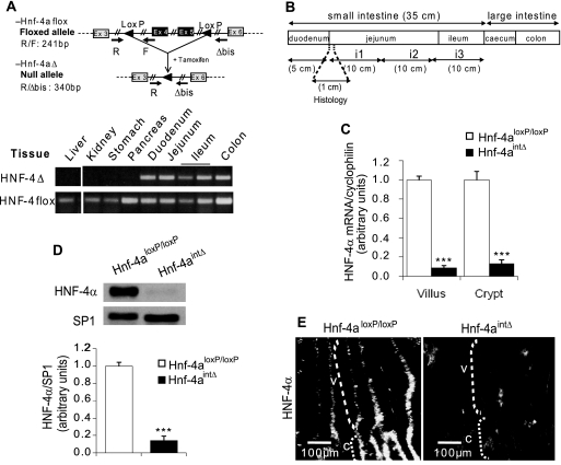
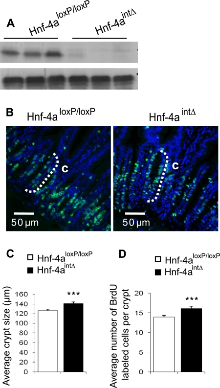
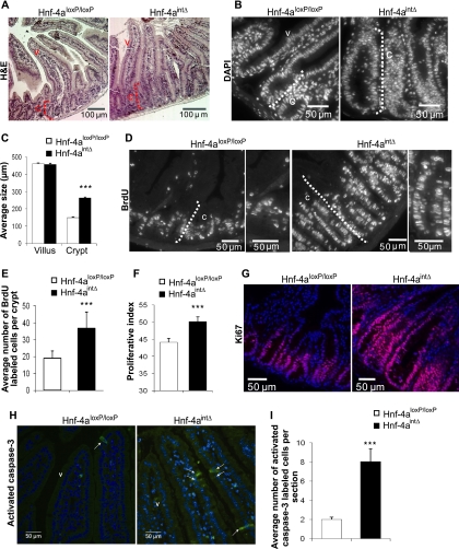
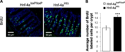
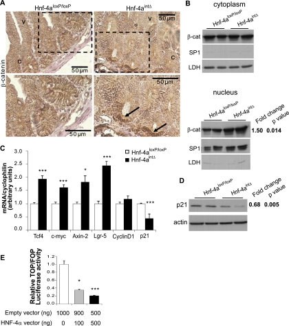
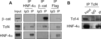
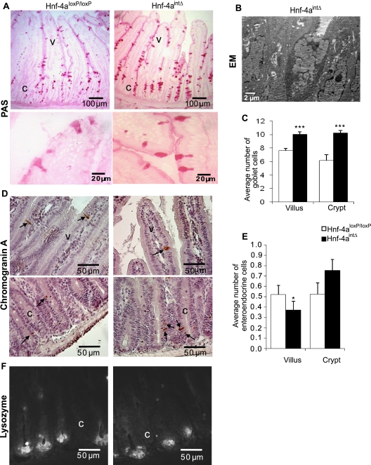
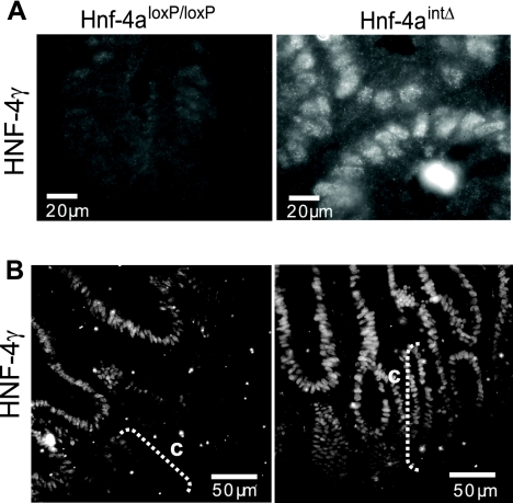
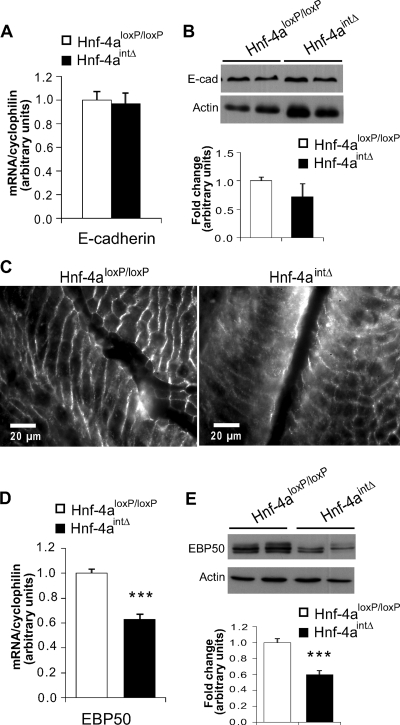
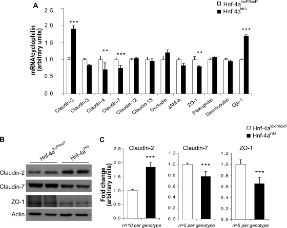
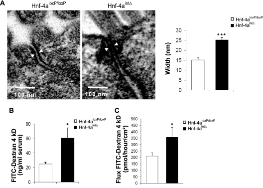
Hepatocyte nuclear factor 4alpha (HNF-4alpha) is a transcription factor which is highly expressed in the intestinal epithelium from duodenum to colon and from crypt to villus. The homeostasis of this constantly renewing epithelium relies on an integrated control of proliferation, differentiation, and apoptosis, as well as on the functional architecture of the epithelial cells. In order to determine the consequences of HNF-4alpha loss in the adult intestinal epithelium, we used a tamoxifen-inducible Cre-loxP system to inactivate the Hnf-4a gene. In the intestines of adult mice, loss of HNF-4alpha led to an increased proliferation in crypts and to an increased expression of several genes controlled by the Wnt/beta-catenin system. This control of the Wnt/beta-catenin signaling pathway by HNF-4alpha was confirmed in vitro. Cell lineage was affected, as indicated by an increased number of goblet cells and an impairment of enterocyte and enteroendocrine cell maturation. In the absence of HNF-4alpha, cell-cell junctions were destabilized and paracellular intestinal permeability increased. Our results showed that HNF-4alpha modulates Wnt/beta-catenin signaling and controls intestinal epithelium homeostasis, cell function, and cell architecture. This study indicates that HNF-4alpha regulates the intestinal balance between proliferation and differentiation, and we hypothesize that it might act as a tumor suppressor.


Figures

FIG. 1.
FIG. 1.
Induction of CreERT2 recombinase leads to specific loss of HNF-4α in the epithelium of the adult mouse small intestine. (A) Schematic representation of the gene with the positions of the three primers used for genotyping (top) and identification of the recombination of the Hnf-4a gene by PCR analysis of genomic DNA from various tissues (bottom). The 340-bp fragment and the 241-bp fragment originate, respectively, from the recombined and intact alleles. The epithelium was obtained by scraping, and the contaminating mesenchyme, where the villin promoter is not active, is at the origin of the 241-bp fragment in the five intestinal samples. Recombination is only detectable in the intestine. (B) Schematic representation of the intestinal cephalocaudal axis. The i1 part of the jejunum was taken to perform RNA and protein analyses. About 1 cm of the proximal jejunum was removed for histological analyses. (C) Quantification of Hnf-4a mRNA levels in epithelial cells from villi and crypts of the jejunum by quantitative RT-PCR. mRNA levels are expressed relative to the amount present in Hnf-4aloxP/loxP mice and were normalized to that of cyclophilin. (D) Representative Western blot analysis and quantification of HNF-4α protein in epithelial cells from the jejunums of control Hnf-4aloxP/loxP and Hnf-4aintΔ mice. The level of HNF-4α is expressed relative to the amount present in Hnf-4aloxP/loxP mice and is normalized to that of SP1. (E) Immunofluorescent staining of HNF-4α in the jejunums of Hnf-4aloxP/loxP and Hnf-4aintΔ mice. Crypts and villi are indicated by the letters C and V, respectively. For each graph, an unpaired Student t test was used to measure significance. ***, P < 0.001. Error bars indicate the standard error of the mean.
FIG. 2.
FIG. 2.
Cell proliferation in adult intestinal epithelium 30 days after tamoxifen injection. (A) Representative Western blot analysis of HNF-4α protein in epithelial cells from the jejunums of control Hnf-4aloxP/loxP and Hnf-4aintΔ mice 30 days following the first injection. (B) BrdU (green) and DAPI (blue) staining in the crypts of jejunums of Hnf-4aintΔ and Hnf-4aloxP/loxP mice. The white dashed bracket delimits the crypt compartment. (C) Average lengths of crypts as determined by measuring 40 crypts from Hnf-4aintΔ and Hnf-4aloxP/loxP mice (n = 3 animals per genotype). (D) Average numbers of BrdU-positive epithelial cells per crypt of Hnf-4aintΔ and Hnf-4aloxP/loxP mice (n = 40 crypts from three animals per genotype).
FIG. 3.
FIG. 3.
Increased cell proliferation and death in the absence of HNF-4α in adult intestinal epithelium. (A) Hematoxylin and eosin (H&E) staining of jejunum sections shows the general structure of the crypt-to-villus axis of Hnf-4aintΔ and Hnf-4aloxP/loxP mice. Note the crypt size of Hnf-4aintΔ mice compared to that of control Hnf-4aloxP/loxP mice. The dashed line indicates the crypt (C), and V marks the villus. (B) DAPI staining to visualize nuclei in the crypts of Hnf-4aintΔ and Hnf-4aloxP/loxP mice. The white dashed bracket delimits the crypt compartment. (C) Representation of the average length of crypts and villi as determined by measuring 40 crypts or villi in each Hnf-4aintΔ or Hnf-4aloxP/loxP mouse (n = 9 animals per genotype). (D) BrdU staining of Hnf-4aintΔ and Hnf-4aloxP/loxP jejunum sections showing the progression rate of cells in S phase. The crypt compartment is delimited by the white dashed bracket. (E) Average number of BrdU-positive epithelial cells per crypt of Hnf-4aintΔ and Hnf-4aloxP/loxP mice (n = 70 crypts from nine animals per genotype). (F) Graphic representation of the proliferative index in crypts of Hnf-4aintΔ and Hnf-4aloxP/loxP mice, i.e., percentage of BrdU-positive epithelial cells per number of crypt cells visualized by DAPI staining. (G) Immunostaining of Ki67 in Hnf-4aintΔ and Hnf-4aloxP/loxP jejunum sections for the identification of epithelial cells undergoing cell cycle progression. (H) Immunostaining of activated caspase 3 in Hnf-4aintΔ and Hnf-4aloxP/loxP jejunum sections for detection of the apoptosis status of epithelial cells. Positive cells are indicated by arrows. (I) Average numbers of activated-caspase 3-positive cells per section of jejunum of Hnf-4aintΔ and Hnf-4aloxP/loxP mice. For each graph, an unpaired Student t test was used to measure significance. ***, P < 0.001. Error bars indicate the standard error of the mean.
FIG. 4.
FIG. 4.
Proliferation in colon epithelium of mice lacking HNF-4α. (A) Representative BrdU staining in the colons of Hnf-4aloxP/loxP and Hnf-4aintΔ mice. (B) Average numbers of BrdU-positive nuclei in epithelia of Hnf-4aloxP/loxP and Hnf-4aintΔ mice. For each graph, an unpaired Student t test was used to measure significance. ***, P < 0.001. Error bars indicate the standard error of the mean.
FIG. 5.
FIG. 5.
Loss of HNF-4α enhances Wnt/β-catenin activity in the adult mouse small intestine. (A) Immunohistochemistry of β-catenin in Hnf-4aintΔ and Hnf-4aloxP/loxP jejunum sections. Membrane and cytosol immunostained β-catenin is detected by a brown precipitate. Sections were counterstained with hematoxylin (blue). Magnifications of the insets (upper panels), delimited by a black dashed square, are shown (lower panels). Cytosolic accumulation of β-catenin is indicated by arrows. Crypts and villi are indicated by C and V, respectively. (B) The level of nuclear β-catenin (β-cat) is enhanced in the absence of HNF-4α. Immunoblot analysis with an anti-β-catenin antibody in crypts of the jejunum revealed the nuclear and cytoplasmic steady-state levels of β-catenin. Cytoplasmic and nuclear fraction purity was assessed through LDH and SP1 immunoblot assays, respectively. The level of protein expression is expressed relative to the amount present in Hnf-4aloxP/loxP mice and is normalized to that of SP1 in the nuclear fraction. Values represent the n-fold change in mean expression between Hnf-4aintΔ and Hnf-4aloxP/loxP mice (n = 3 per genotype). (C) Quantitative RT-PCRs for Wnt target gene expression in adult Hnf-4aintΔ and Hnf-4aloxP/loxP jejunum sections. Significant upregulation of Tcf-4, axin 2, c-Myc, and Lgr-5 and significant downregulation of p21 were seen after HNF-4α loss. The mRNA levels are expressed relative to the amount present in Hnf-4aloxP/loxP mice and were normalized to the cyclophilin mRNA level. (D) The p21 level is regulated by HNF-4α in the adult jejunum. Immunoblot analysis with an anti-p21 antibody revealed the steady-state levels of p21 and actin (loading control). The value represents the n-fold change in the level of p21 expression between Hnf-4aintΔ and Hnf-4aloxP/loxP mice and was normalized to that of actin. (E) Overexpression of HNF-4α in HCT116 cells inhibits TOPflash activity. HCT116 cells were transfected with TOPflash, a β-catenin/TCF/LEF-responsive luciferase reporter plasmid containing TCF-4-binding sites, or the corresponding negative control FOPflash, along with HNF-4α and β-galactosidase vectors. Luciferase activity was normalized to the corresponding β-galactosidase activity to obtain the relative TOP/FOP luciferase activity. The graph represents the average luciferase activity (n = 3 independent experiments performed in triplicate). For each graph, an unpaired Student t test was used to measure significance. ***, **, and *, P < 0.001, P < 0.01, and P < 0.05, respectively. Error bars indicate the standard error of the mean.
FIG. 6.
FIG. 6.
HNF-4α interacts with Tcf-4 in intestinal epithelial cells. Coimmunoprecipitation analysis of HNF-4α, Tcf-4, and β-catenin (β-cat). (A) Lysates from HCT116 cells transfected with plasmids encoding HNF-4α and Flag-Tcf4 were subjected to immunoprecipitation with anti-HNF-4α, anti-Flag, or anti-β-catenin antibodies as indicated at the top. The input (4% of the total cell extracts) and specific (IP) and nonspecific (IgG) immunoprecipitates were analyzed by Western blotting with the indicated antibodies. (B) Western blot detection of endogenous HNF-4α in Caco-2/TC7 cells after immunoprecipitation with the indicated antibodies.
FIG. 7.
FIG. 7.
Altered number of goblet cells in the small intestines of adult Hnf-4aintΔ mice. (A) PAS (32) staining (pink) of goblet cells in adult Hnf-4aintΔ and Hnf-4aloxP/loxP jejunums. (B) Electron microscopy showing a mature goblet cell in adult Hnf-4aintΔ jejunum. Optical microscopy shows that the goblet cells are open at the luminal side, and electron microscopy shows that the secretory granules occupy a large portion of the cytoplasm. (C) Average number of PAS-positive jejunum cells of adult Hnf-4aintΔ and Hnf-4aloxP/loxP mice per villus or per crypt (n = 8 crypt-to-villus axes and 3 mice per genotype). (D) Chromogranin A immunostaining (brown) and hematoxylin (blue) counterstaining of adult Hnf-4aintΔ and Hnf-4aloxP/loxP jejunum sections to visualize enteroendocrine cells. Arrows indicate specific staining of enteroendocrine cells. (E) Average number of chromogranin A-positive cells per villus or per crypt of adult Hnf-4aintΔ and Hnf-4aloxP/loxP mouse jejunum (n = 40 crypt-to-villus axes from eight animals per genotype). (F) Immunostaining of lysozyme of adult Hnf-4aintΔ and Hnf-4aloxP/loxP mouse jejunum sections to visualize Paneth cells. Crypts and villi are indicated by C and V, respectively. For each graph, an unpaired Student t test was used to measure significance. *** and *, P < 0.001 and P < 0.05, respectively. Error bars indicate the standard error of the mean.
FIG. 8.
FIG. 8.
Expression of HNF-4γ in the absence of HNF-4α. Representative HNF-4γ staining observed in jejunum sections of Hnf-4aloxP/loxP and Hnf-4aintΔ mice 8 (A) and 30 (B) days after the first tamoxifen injection.
FIG. 9.
FIG. 9.
Expression of E-cadherin and EBP50 in the absence of HNF-4α. (A) The E-cadherin mRNA level from the villus epithelium was analyzed by quantitative RT-PCR in Hnf-4aloxP/loxP and Hnf-4aintΔ mice. The mRNA level is expressed relative to the amount present in Hnf-4aloxP/loxP mice and is normalized to that of cyclophilin. (B) Representative Western blot analysis of E-cadherin in the villus epithelia of Hnf-4aloxP/loxP and Hnf-4aintΔ mice. The histogram represents the quantification of the level of E-cadherin (E-cad) protein (n = 5 per genotype). The level of E-cadherin expression is expressed relative to the amount present in Hnf-4aloxP/loxP mice and is normalized to that of actin. (C) Immunostaining of E-cadherin in the jejunums of Hnf-4aloxP/loxP and Hnf-4aintΔ mice. Note the diffuse staining of E-cadherin in Hnf-4aintΔ mice. (D) The EBP50 mRNA level from the villus epithelium was analyzed by real-time quantitative PCR in Hnf-4aloxP/loxP and Hnf-4aintΔ mice. The mRNA level is expressed relative to the amount present in Hnf-4aloxP/loxP mice and is normalized to that of cyclophilin. (E) Representative Western blot analysis of EBP50 in the villus epithelia of Hnf-4aloxP/loxP and Hnf-4aintΔ mice. The histogram represents the quantification of the level of EBP50 protein (n = 7 per genotype). The level of EBP50 expression is expressed relative to the amount present in Hnf-4aloxP/loxP mice and is normalized to that of actin expression. For each graph, an unpaired Student t test was used to measure significance. ***, P < 0.001. Error bars indicate the standard error of the mean.
FIG. 10.
FIG. 10.
Expression of tight junction-associated proteins in the absence of HNF-4α. (A) The levels of mRNAs encoding different junction proteins from the villus epithelium were analyzed by quantitative RT-PCR in Hnf-4aloxP/loxP and Hnf-4aintΔ mice. mRNA levels are expressed relative to those of Hnf-4aloxP/loxP mice and are normalized to the cyclophilin mRNA level. (B) Representative Western blot analysis of claudin 2, claudin 7, and ZO-1 in the jejunums of Hnf-4aloxP/loxP and Hnf-4aintΔ mice. (C) Quantification of the expression of tight junction-associated proteins by scanning Western blot analysis. The level of protein expression is expressed relative to the amount present in Hnf-4aloxP/loxP mice and is normalized to that of actin. Values represent the n-fold changes in mean expression between Hnf-4aintΔ and Hnf-4aloxP/loxP mice. For each graph, an unpaired Student t test was used to measure significance. *** and **, P < 0.001 and P < 0.01, respectively. Error bars indicate the standard error of the mean.
FIG. 11.
FIG. 11.
Distension of tight junctions and increased paracellular permeability in the absence of HNF-4α. (A) Electron microscopic images showing the intercellular space in tight junctions of Hnf-4aloxP/loxP and Hnf-4aintΔ mice. The histogram represents the average width of tight junctions, measured between the two external membranes, as indicated by arrowheads (8 to 12 junctions and three animals per genotype). (B) In vivo paracellular permeability analysis done by measuring the FITC-dextran in serum samples from Hnf-4aloxP/loxP and Hnf-4aintΔ mice (n = 9 animals per genotype). (C) Paracellular permeability analysis done by measuring FITC-dextran flux in Ussing chambers with Hnf-4aloxP/loxP and Hnf-4aintΔ mouse intestine sections (n = 12 animals per genotype.). For each graph, an unpaired Student t test was used to measure significance. *** and *, P < 0.001 and P < 0.05, respectively. Error bars indicate the standard error of the mean.

References

    1. Ahn, S. H., Y. M. Shah, J. Inoue, K. Morimura, I. Kim, S. Yim, G. Lambert, R. Kurotani, K. Nagashima, F. J. Gonzalez, and Y. Inoue. 2008. Hepatocyte nuclear factor 4α in the intestinal epithelial cells protects against inflammatory bowel disease. Inflamm. Bowel Dis. 14:908-920. - PMC - PubMed
    1. Archer, A., D. Sauvaget, V. Chauffeton, P. E. Bouchet, J. Chambaz, M. Pincon-Raymond, P. Cardot, A. Ribeiro, and M. Lacasa. 2005. Intestinal apolipoprotein A-IV gene transcription is controlled by two hormone-responsive elements: a role for hepatic nuclear factor-4 isoforms. Mol. Endocrinol. 19:2320-2334. - PubMed
    1. Babeu, J. P., M. Darsigny, C. R. Lussier, and F. Boudreau. 2009. Hepatocyte nuclear factor 4α contributes to an intestinal epithelial phenotype in vitro and plays a partial role in mouse intestinal epithelium differentiation. Am. J. Physiol. Gastrointest. Liver Physiol. 297:G124-G134. - PubMed
    1. Barreau, F., U. Meinzer, F. Chareyre, D. Berrebi, M. Niwa-Kawakita, M. Dussaillant, B. Foligne, V. Ollendorff, M. Heyman, S. Bonacorsi, T. Lesuffleur, G. Sterkers, M. Giovannini, and J. P. Hugot. 2007. CARD15/NOD2 is required for Peyer's patches homeostasis in mice. PLoS ONE 2:e523. - PMC - PubMed
    1. Battle, M. A., G. Konopka, F. Parviz, A. L. Gaggl, C. Yang, F. M. Sladek, and S. A. Duncan. 2006. Hepatocyte nuclear factor 4α orchestrates expression of cell adhesion proteins during the epithelial transformation of the developing liver. Proc. Natl. Acad. Sci. USA 103:8419-8424. - PMC - PubMed

Publication types