. 2009 Dec;77(12):5272-80.
doi: 10.1128/IAI.00201-09. Epub 2009 Oct 5.

The Sca2 autotransporter protein from Rickettsia conorii is sufficient to mediate adherence to and invasion of cultured mammalian cells

Affiliations

The Sca2 autotransporter protein from Rickettsia conorii is sufficient to mediate adherence to and invasion of cultured mammalian cells

Marissa M Cardwell et al. Infect Immun. 2009 Dec.

Abstract

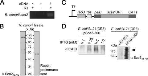
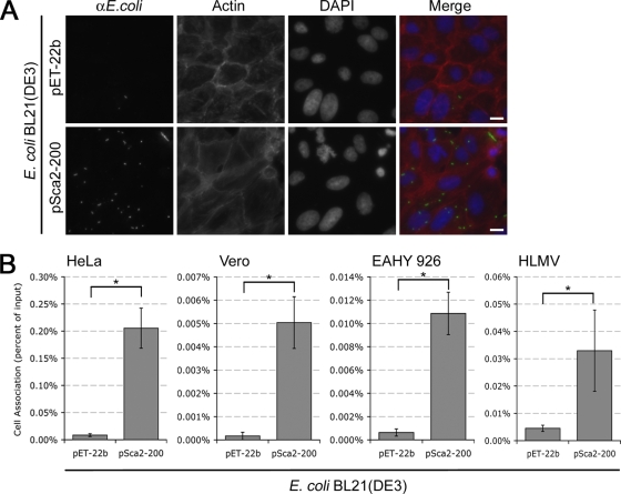
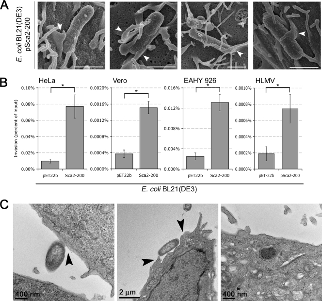
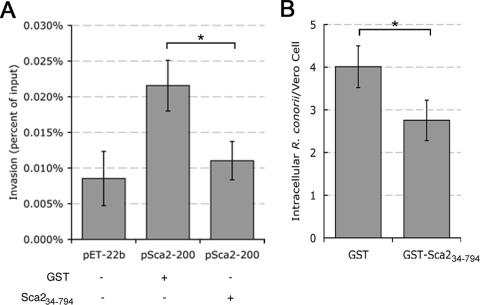
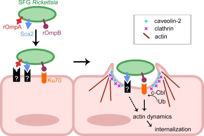
Obligate intracellular bacteria of the genus Rickettsia must adhere to and invade the host endothelium in order to establish an infection. These processes require the interaction of rickettsial surface proteins with mammalian host cell receptors. A previous bioinformatic analysis of sequenced rickettsial species identified a family of at least 17 predicted "surface cell antigen" (sca) genes whose products resemble autotransporter proteins. Two members of this family, rOmpA and rOmpB of spotted fever group (SFG) rickettsiae have been identified as adhesion and invasion factors, respectively; however, little is known about the putative functions of the other sca gene products. An intact sca2 gene is found in the majority of pathogenic SFG rickettsiae and, due to its sequence conservation among these species, we predict that Sca2 may play an important function at the rickettsial surface. Here we have shown that sca2 is transcribed and expressed in Rickettsia conorii and have used a heterologous gain-of-function assay in E. coli to determine the putative role of Sca2. Using this system, we have demonstrated that expression of Sca2 at the outer membrane of nonadherent, noninvasive E. coli is sufficient to mediate adherence to and invasion of a panel of mammalian cells, including endothelial cells. Furthermore, soluble Sca2 protein is capable of diminishing R. conorii invasion of cultured mammalian cells. This is the first evidence that Sca2 participates in the interaction between SFG rickettsiae and host cells and suggests that in addition to other surface proteins, Sca2 may play a critical role in rickettsial pathogenesis.


Figures

FIG. 1.
FIG. 1.
Detection of endogenous Sca2 protein in R. conorii and expression of recombinant Sca2 protein in E. coli. (A) Agarose gel electrophoresis analysis of PCR products resulting from amplification of R. conorii cDNA using a sca2-specific primer pair (Table 2). PCR product is 240 bp in size. A no-template control (-cDNA) and no-reverse-transcriptase control (-RT) are included to control for environmental DNA contamination and genomic DNA contamination, respectively. (B) Western immunoblot analysis of R. conorii whole-cell lysate probed with rabbit anti-Sca234-794 hyperimmune sera (left panel) and rabbit preimmune sera (right panel). (C) Schematic of the pET-22b expression vector with the inserted sca2 open reading frame. Relevant features of the 5′ and 3′ ends of pSca2-200, including the in-frame N-terminal pelB leader sequence and the C-terminal His6 tag are shown. (D) Western immunoblot analysis of outer membrane fractions of E. coli BL21(DE3) expressing the empty vector (pET-22b) or full-length R. conorii Sca2 (pSca2-200) probed with anti-His6 (left panel) and anti-Sca234-794 antibodies (right panel). Maximal expression of recombinant Sca2 is observed with 0.5 mM IPTG induction (left panel), resulting in a protein product larger than 170 kDa (arrow).
FIG. 2.
FIG. 2.
Expression of Sca2 in E. coli is sufficient to mediate adherence to mammalian cells. (A) Fluorescence micrographs of monolayers of Vero cells infected with E. coli BL21(DE3) expressing the empty vector (pET-22b) or the full-length Sca2 protein from R. conorii (pSca2-200). Confluent monolayers of Vero cells were infected for 20 min at 37°C, washed repeatedly with PBS, and then processed for immunofluorescence. Scale bars, 10 μm. (B) CFU-based quantification of bacterial adherence to mammalian host cells. HeLa, Vero, EAHY 926, and HLMV cells were infected with induced cultures as in panel A. Confluent monolayers were infected with induced bacteria for 20 min at 37°C and washed repeatedly with PBS, and then cell-associated bacteria were extracted from host cells by detergent lysis, and plated for CFU quantification. Association was determined as the % CFU of cell-associated bacteria from the initial bacterial inoculums. Actual percentages varied from assay to assay (ranging from 0.001 to 0.2%) depending on the passage number of cells used and the expression of Sca2 at the E. coli outer membrane. *, P < 0.05. The data presented are representative of at least three independent assays for each cell line. Error bars represent the standard deviation of each data set.
FIG. 3.
FIG. 3.
Sca2 mediates invasion of mammalian cells. (A) Scanning electron micrographs examining the surface interaction of Sca2-expressing E. coli with HeLa cells. HeLa cell monolayers on glass coverslips were infected with induced bacteria for 20 min and then processed for SEM. White arrowheads highlight possible Sca2-mediated cellular membrane rearrangements. Scale bars, 1 μm. (B) CFU-based quantification of bacterial invasion of HeLa, Vero, EAHY 926, and HLMV cells. Confluent cell monolayers were infected for 1 h with E. coli BL21(DE3) expressing the empty vector (pET-22b) or Sca2 from R. conorii (pSca2-200) and assessed for invasion by gentamicin protection assay. Invasion is presented as the percentage of bacteria recovered after the gentamicin challenge out of the inoculums. Actual percentages varied from assay to assay (ranging from 0.0007 to 0.07%), depending on the passage number of the cells used and the expression of Sca2 at the E. coli outer membrane. *, P < 0.05. The data presented are representative of at least two independent experiments for each individual cell line. Error bars represent the standard deviation of each data set. (C) Transmission electron micrographs of HeLa cells infected with Sca2-expressing E. coli demonstrate different steps of the uptake process, including the initial attachment event (left panel), the induction of changes in the plasma membrane (arrows in middle panel), and the presence of bacteria within membrane bound vacuoles (right panel). Scale bars are indicated in each panel.
FIG. 4.
FIG. 4.
Soluble Sca2 protein inhibits rickettsial invasion. (A) Monolayers of HeLa cells were incubated with 1 μM soluble GST or GST-Sca234-794 protein for 20 min at 37°C and 5%CO2 prior to infection with. E. coli BL21(DE3) expressing pSca2-200. BL21(DE3) harboring pET-22b was used as a negative control. Bacterial invasion was assessed as described in Fig. 3B. (B) Vero cell monolayers seeded at 90% confluence were incubated with 8 μM GST or GST-Sca234-794 protein prior to infection with R. conorii for 30 min. Total rickettsial association and intracellular bacteria were enumerated by differential immunofluorescence microscopy. *, P < 0.05. The data presented are representative of at least two independent experiments. The error bars represent the standard deviation from the average in these experiments.
FIG. 5.
FIG. 5.
Model of SFG Rickettsia interactions with mammalian cells. Interactions of SFG Rickettsia with host cells are likely governed by coordinated interactions between rOmpB-Ku70 and Sca2 with an unknown mammalian receptor (black box). Scanning and transmission electron microscopy analyses revealed similarities between rOmpB and Sca2-mediated uptake of mammalian cells, suggesting that while signals may be triggered by different receptors at the plasma membrane, signals ultimately converge to recruit localized actin filaments and components of the endocytic machinery (clathrin, caveolin-2, and c-Cbl) to entry foci.

References

    1. Blanc, G., M. Ngwamidiba, H. Ogata, P. E. Fournier, J. M. Claverie, and D. Raoult. 2005. Molecular evolution of Rickettsia surface antigens: evidence of positive selection. Mol. Biol. Evol. 20:2073-2083. - PubMed
    1. Chan, Y. G. Y., M. M. Cardwell, T. M. Hermanas, T. Uchiyama, and J. J. Martinez. 2009. Rickettsial outer-membrane protein B (rOmpB) mediates bacterial invasion through Ku70 in an actin, c-Cbl, clathrin and caveolin 2-dependent manner. Cell. Microbiol. 11:629-644. - PMC - PubMed
    1. Cossart, P., and P. J. Sansonetti. 2004. Bacterial invasion: the paradigms of enteroinvasive pathogens. Science 304:242-248. - PubMed
    1. Edgell, C. J., C. C. McDonald, and J. B. Graham. 1983. Permanent cell line expressing human factor VIII-related antigen established by hybridization. Proc. Natl. Acad. Sci. USA 80:3734-3737. - PMC - PubMed
    1. Feng, H. M., T. Whitworth, J. P. Olano, V. L. Popov, and D. H. Walker. 2004. Fc-dependent polyclonal antibodies and antibodies to outer membrane proteins A and B, but not to lipopolysaccharide, protect SCID mice against fatal Rickettsia conorii infection. Infect. Immun. 72:2222-2228. - PMC - PubMed

Publication types