. 2009 Dec 4;284(49):34390-9.
doi: 10.1074/jbc.M109.021873. Epub 2009 Oct 5.

Bax activates endophilin B1 oligomerization and lipid membrane vesiculation

Affiliations

Bax activates endophilin B1 oligomerization and lipid membrane vesiculation

Tatiana K Rostovtseva et al. J Biol Chem. .

Abstract

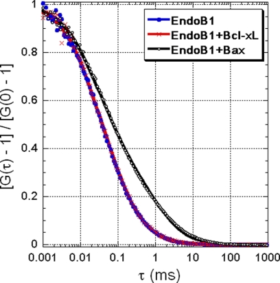
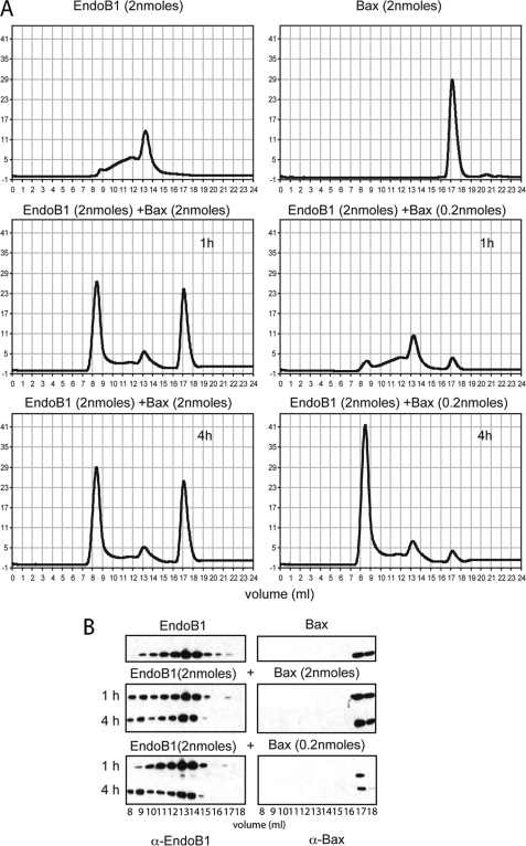
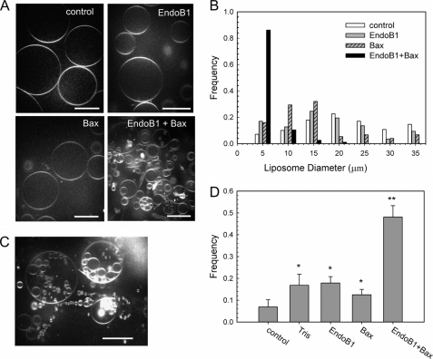
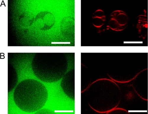
Endophilins participate in membrane scission events that occur during endocytosis and intracellular organelle biogenesis through the combined activity of an N-terminal BAR domain that interacts with membranes and a C-terminal SH3 domain that mediates protein binding. Endophilin B1 (Endo B1) was identified to bind Bax, a Bcl-2 family member that promotes apoptosis, through yeast two-hybrid protein screens. Although Endo B1 does not bind Bax in healthy cells, during apoptosis, Endo B1 interacts transiently with Bax and promotes cytochrome c release from mitochondria. To explore the molecular mechanism of action of Endo B1, we have analyzed its interaction with Bax in cell-free systems. Purified recombinant Endo B1 in solution displays a Stokes radius indicating a tetrameric quarternary structure. However, when incubated with purified Bax, it assembles into oligomers more than 4-fold greater in molecular weight. Although Endo B1 oligomerization is induced by Bax, Bax does not stably associate with the high molecular weight Endo B1 complex. Endo B1 oligomerization requires its C-terminal Src homology 3 domain and is not induced by Bcl-xL. Endo B1 combined with Bax reduces the size and changes the morphology of giant unilamellar vesicles by inducing massive vesiculation of liposomes. This activity of purified Bax protein to induce cell-free assembly of Endo B1 may reflect its activity in cells that regulates apoptosis and/or mitochondrial fusion.


Figures

FIGURE 1.
FIGURE 1.
Oligomerization of Endo B1 in the presence of Bax. Normalized FCS correlation functions show a time shift of the correlation of Endo B1 plus Bax solution with respect to that of Endo B1 or Endo B1 plus Bcl-xL, indicating oligomerization of Endo B1 in the presence of Bax. The solid lines are fits of the data, where a two-component model is used (Equation 4). Each curve is labeled with the components in the solutions.
FIGURE 2.
FIGURE 2.
Bax induces oligomerization of Endo B1. Gel filtration profiles of Endo B1 or Endo B1ΔC before and after incubation for 1 h with Bax (A) or Bcl-xL (B). Gel filtration of Bax and Bcl-xL alone was performed for comparison of their elution times (A and B). Fractions 8–18 from the Endo B1, Bax, and Endo B1 plus Bax gel filtration columns were analyzed by Western blot using anti-Bax and anti-Endo B1 antibodies (C).
FIGURE 3.
FIGURE 3.
Substoichiometric levels of Bax oligomerize Endo B1. A, gel filtration profiles of Endo B1 (2 nmol) incubated with Bax (2 nmol) or with Bax (0.2 nmol) for 1 and 4 h as well as Bax and Endo B1 alone. Fractions 8–18 from the Endo B1 alone, Bax alone, and Endo B1 (2 nmol) incubated with Bax (2 nmol or 0.2 nmol) gel filtration columns were analyzed by Western blot using anti-Bax and anti-Endo B1 antibodies (B).
FIGURE 4.
FIGURE 4.
Endo B1 with Bax induces massive vesiculation and liposome size reduction. A, control image before protein addition (upper left). Endo B1 or Bax does not induce liposome shape deformation (upper right and lower left, respectively). The sphericity of these GUVs indicates a lack of osmotic stress. Confocal images were taken 30 min after the addition of 490 nm Endo B1 or Bax. Endo B1 preincubated with Bax affects shape and size of GUVs (lower right). The image was taken 20 min after the Endo B1 plus Bax (1:1 mol/mol) addition. Scale bars here and elsewhere, 20 μm. B, size distribution of GUVs obtained before and after the addition of Endo B1 plus Bax, Endo B1, or Bax. Normalized number of GUVs is plotted versus their size. Data are the summary of the analysis of at least three independent experiments for each condition with multiple images taken in each experiment. C, a representative image of large GUVs with entrapped multiple smaller vesicles. D, number of GUVs with smaller vesicle inclusions increases after the addition of Endo B1 plus Bax. A summary of the statistical analysis of the percentage of GUVs with inclusions before and after the addition of Endo B1 plus Bax, Endo B1, Bax, or corresponding aliquots of Tris buffer as a control. The number of liposomes analyzed per experiment was ∼150. Statistical analysis was done using a two-tailed t test (a = 0.05) as a comparison with control after the addition of 0.2 mm Tris buffer (**, p < 0.04) and as a comparison with control before protein injection (*, p < 0.1).
FIGURE 5.
FIGURE 5.
Internal small vesicles appear to bud off from the GUV membrane. A, images of GUVs generated after sequential addition of Alexa-488 and unlabeled Endo B1 preincubated with Bax. Free dye fills some of the small vesicles (green circles) inside the larger one without dye (empty dark green circles). B, confocal images of GUVs in the presence of free Alexa-488 (green) show that dye does not penetrate through the membrane of GUV (up to 2 h). Images at the left were taken at the 488-nm excitation wave and show Alexa-488. The same images at the right were taken at 568 nm and show liposome membrane labeled with rhodamine-dioleoylphosphatidylethanolamine (red).

Similar articles

Cited by

References

    1. Farsad K., Ringstad N., Takei K., Floyd S. R., Rose K., De Camilli P. (2001) J. Cell Biol. 155, 193–200 - PMC - PubMed
    1. Cuddeback S. M., Yamaguchi H., Komatsu K., Miyashita T., Yamada M., Wu C., Singh S., Wang H. G. (2001) J. Biol. Chem. 276, 20559–20565 - PubMed
    1. Pierrat B., Simonen M., Cueto M., Mestan J., Ferrigno P., Heim J. (2001) Genomics 71, 222–234 - PubMed
    1. Takahashi Y., Karbowski M., Yamaguchi H., Kazi A., Wu J., Sebti S. M., Youle R. J., Wang H. G. (2005) Mol. Cell Biol. 25, 9369–9382 - PMC - PubMed
    1. Karbowski M., Jeong S. Y., Youle R. J. (2004) J. Cell Biol. 166, 1027–1039 - PMC - PubMed

Publication types