. 2009 Oct 20;120(16):1606-15.
doi: 10.1161/CIRCULATIONAHA.109.872200. Epub 2009 Oct 5.

Activin A and follistatin-like 3 determine the susceptibility of heart to ischemic injury

Affiliations

Activin A and follistatin-like 3 determine the susceptibility of heart to ischemic injury

Yuichi Oshima et al. Circulation. .

Abstract

Background: Transforming growth factor-beta family cytokines have diverse actions in the maintenance of cardiac homeostasis. Activin A is a member of this family whose regulation and function in heart are not well understood at a molecular level. Follistatin-like 3 (Fstl3) is an extracellular regulator of activin A protein, and its function in the heart is also unknown.

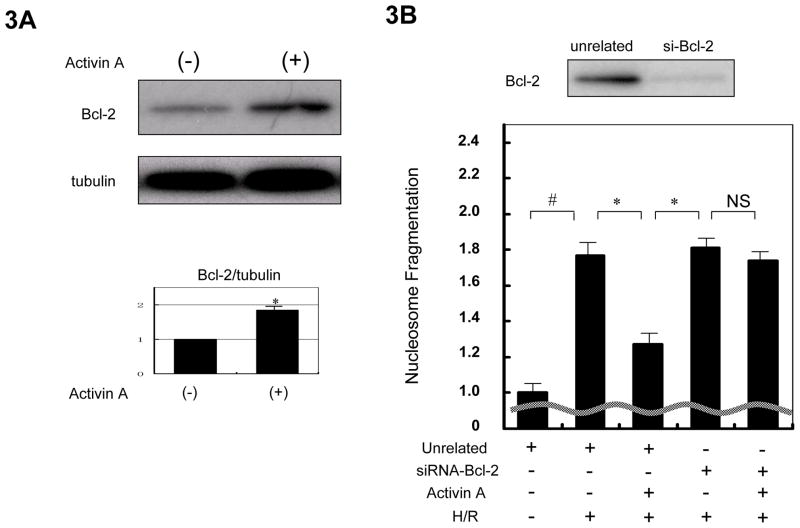
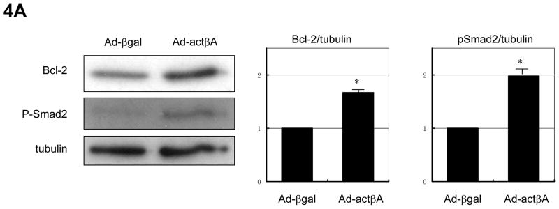
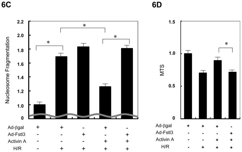
Methods and results: We analyzed the expression of various transforming growth factor-beta superfamily cytokines and their binding partners in mouse heart. Activin betaA and Fstl3 were upregulated in models of myocardial injury. Overexpression of activin A with an adenoviral vector (Ad-actbetaA) or treatment with recombinant activin A protein protected cultured myocytes from hypoxia/reoxygenation-induced apoptosis. Systemic overexpression of activin A in mice by intravenous injection of Ad-actbetaA protected hearts from ischemia/reperfusion injury. Activin A induced the expression of Bcl-2, and ablation of Bcl-2 by small interfering RNA abrogated its protective action in myocytes. The protective effect of activin A on cultured myocytes was abolished by treatment with Fstl3 or by a pharmacological activin receptor-like kinase inhibitor. Cardiac-specific Fstl3 knockout mice showed significantly smaller infarcts after ischemia/reperfusion injury that was accompanied by reduced apoptosis.

Conclusions: Activin A and Fstl3 are induced in heart by myocardial stress. Activin A protects myocytes from death, and this activity is antagonized by Fstl3. Thus, the relative expression levels of these factors after injury is a determinant of cell survival in the heart.


Figures

Figure 1
Figure 1
Fstl3 and Activin βA are specifically induced after cardiac injury. Expression analysis of Activin βA, Fstl3, Follistatin, and Inhibinα in murine models of myocardial infarction (MI) (A), transverse aortic constriction (TAC) (B) and ischemia/reperfusion (I/R) (C). In the MI model, samples were taken separately from ischemic zone (infarct area) and non-ischemic zone (remote area) three days after the onset of MI. For the pressure overload model, samples were taken 7 days after TAC surgery. QRT-PCR was performed to determine the mRNA level of each transcript and the data were compared to the GAPDH level and normalized to the mean value of controls. n=4−6. *P<0.05 vs. sham and #P<0.01 vs. sham. (D) Upregulation of Activin A protein following myocardial infarction. The upper panel is a western blot analysis for Activin A performed under non-reducing condition and the lower panel is a blot for α-tubulin using the same samples under reducing conditions. The histogram shows the quantification of the band intensities for Activin A compared to that of tubulin. #P<0.01 vs. sham. (E) Upregulation of Activin A and Fstl3 proteins after H/R treatment in NRVM cultures. Representative images of immunoblots of the culture media, 24 hrs after addition to cells, and the cell pellet lysates.
Figure 1
Figure 1
Fstl3 and Activin βA are specifically induced after cardiac injury. Expression analysis of Activin βA, Fstl3, Follistatin, and Inhibinα in murine models of myocardial infarction (MI) (A), transverse aortic constriction (TAC) (B) and ischemia/reperfusion (I/R) (C). In the MI model, samples were taken separately from ischemic zone (infarct area) and non-ischemic zone (remote area) three days after the onset of MI. For the pressure overload model, samples were taken 7 days after TAC surgery. QRT-PCR was performed to determine the mRNA level of each transcript and the data were compared to the GAPDH level and normalized to the mean value of controls. n=4−6. *P<0.05 vs. sham and #P<0.01 vs. sham. (D) Upregulation of Activin A protein following myocardial infarction. The upper panel is a western blot analysis for Activin A performed under non-reducing condition and the lower panel is a blot for α-tubulin using the same samples under reducing conditions. The histogram shows the quantification of the band intensities for Activin A compared to that of tubulin. #P<0.01 vs. sham. (E) Upregulation of Activin A and Fstl3 proteins after H/R treatment in NRVM cultures. Representative images of immunoblots of the culture media, 24 hrs after addition to cells, and the cell pellet lysates.
Figure 2
Figure 2
Activin A protects cardiac myocytes from H/R induced injury. (A) NRVMs were pretreated with different concentrations of Activin A for 8 hours before the exposure to 12 hours of hypoxia followed by 24 hours of reoxygenation (H/R). Cell viability was determined by the MTS assay. Apoptosis indicated by nucleosome fragmentation assay (B) and caspase-3 and -7 activities (C) were measured in NRVMs pretreated with 25ng/mL of Activin A before exposure to H/R. (D) Thirty minutes before addition of Activin A, NRVMs were pretreated with or without the inhibitor SB431542. Cell viability was measured by MTS assay after H/R treatment. *P<0.05 and #P<0.01.
Figure 2
Figure 2
Activin A protects cardiac myocytes from H/R induced injury. (A) NRVMs were pretreated with different concentrations of Activin A for 8 hours before the exposure to 12 hours of hypoxia followed by 24 hours of reoxygenation (H/R). Cell viability was determined by the MTS assay. Apoptosis indicated by nucleosome fragmentation assay (B) and caspase-3 and -7 activities (C) were measured in NRVMs pretreated with 25ng/mL of Activin A before exposure to H/R. (D) Thirty minutes before addition of Activin A, NRVMs were pretreated with or without the inhibitor SB431542. Cell viability was measured by MTS assay after H/R treatment. *P<0.05 and #P<0.01.
Figure 3
Figure 3
Cytoprotection by Activin A is mediated by upregulation of the anti-apoptotic protein Bcl-2. (A) Activin A induced Bcl-2 expression in NRVMs. A representative immunoblot is shown. Blots for α-tubulin were performed to indicate the equal loading. The histogram shows quantification of the band intensities to indicate a statistically a significant increase in Bcl-2 protein expression following treatment with Activin A. *P<0.05. (B) Ablation of Bcl-2 expression blocks the cytoprotection conferred by Activin A. The upper panel shows a representative western blot assessing the efficiency of siRNA targeting Bcl-2. The lower panel displays the effect of Bcl-2 knock-down on Activin A-mediated cytoprotection of NRVMs as determined by nucleosome fragmentation assay. Apoptosis was induced by hypoxia/reoxygenation treatment.*P<0.05 and #P<0.01.
Figure 4
Figure 4
Adenovirus-encoded Activin A induces Bcl-2 and protects cardiac myocytes from stress. (A) Representative immunoblot images showing that transfection of adenoviral vector expressing Activin βA (Ad-actβA) at an MOI of 50 resulted in increased expression of Bcl-2 and phosphorylation of Smad2. Membranes were blotted for α-tubulin to indicate equal protein loading. Histogram show quantification of the band intensities. *P<0.05. Transduction of Ad-actβA (MOI=50) reduced apoptosis, assessed by the nucleosome fragmentation assay (B), and preserved cell viability, assessed by the MTS assay, (C) against H/R stress. An adenovirus vector expressing β-galactosidase (Ad-βgal) was used as a control in these experiments at an MOI of 50. *P<0.05 and #P<0.01.
Figure 4
Figure 4
Adenovirus-encoded Activin A induces Bcl-2 and protects cardiac myocytes from stress. (A) Representative immunoblot images showing that transfection of adenoviral vector expressing Activin βA (Ad-actβA) at an MOI of 50 resulted in increased expression of Bcl-2 and phosphorylation of Smad2. Membranes were blotted for α-tubulin to indicate equal protein loading. Histogram show quantification of the band intensities. *P<0.05. Transduction of Ad-actβA (MOI=50) reduced apoptosis, assessed by the nucleosome fragmentation assay (B), and preserved cell viability, assessed by the MTS assay, (C) against H/R stress. An adenovirus vector expressing β-galactosidase (Ad-βgal) was used as a control in these experiments at an MOI of 50. *P<0.05 and #P<0.01.
Figure 5
Figure 5
Adenovirus-mediated overexpression of Activin A protects the heart from ischemia/reperfusion (I/R) injury. (A) Representative western blot analysis, performed under non-reducing conditions, of plasma samples collected three days after injection of Ad-βgal or Ad-actβA. Histogram shows quantification of infarct area induced by I/R three days after adenoviral injection. *P<0.05. (B) Representative images of myocardium stained with TUNEL (green) and sarcomeric actin (red) (upper panels), and merged with DAPI (blue) (lower panels). Histogram shows quantification of TUNEL-positive cells in the myocardium after I/R. *P<0.05.
Figure 6
Figure 6
Fstl3 inhibits Activin A action in NRVMs. (A, B) NRVMs, transduced with Ad-Fstl3 (50MOI) or Ad-Fstl1 (50MOI), were stimulated with 25 ng/mL of recombinant Activin A for indicated periods of time, and phosphorylation of Smad2 was determined by western blot analysis. Immunoblots for α-tubulin were performed as a loading control. (C, D) NRVMs were transduced with Ad-Fstl3 or Ad-βgal at an MOI of 50 (C) or 10 (D) and exposed to H/R treatment in the presence or absence of pretreatment with 25ng/mL of Activin A. Apoptosis was examined by nucleosome fragmentation assay (C) and cell viability was assessed by the MTS assay (D). *P<0.05.
Figure 6
Figure 6
Fstl3 inhibits Activin A action in NRVMs. (A, B) NRVMs, transduced with Ad-Fstl3 (50MOI) or Ad-Fstl1 (50MOI), were stimulated with 25 ng/mL of recombinant Activin A for indicated periods of time, and phosphorylation of Smad2 was determined by western blot analysis. Immunoblots for α-tubulin were performed as a loading control. (C, D) NRVMs were transduced with Ad-Fstl3 or Ad-βgal at an MOI of 50 (C) or 10 (D) and exposed to H/R treatment in the presence or absence of pretreatment with 25ng/mL of Activin A. Apoptosis was examined by nucleosome fragmentation assay (C) and cell viability was assessed by the MTS assay (D). *P<0.05.
Figure 7
Figure 7
Fstl3 ablation in heart. (A,B) QRT-PCR was carried out to evaluate Fslt3 mRNA expression in hearts of wild-type (W) or αMHC-Cre (Cre) mice crossed with Fstl3flox/flox mice using cDNA produced from whole heart extracts (A) or isolated cardiac myocytes (B). *P<0.05. (C) Heart weight/body weight ratio (HW/BW) in eight week old male mice. (D, E) Representative images of immunoblots of Bcl-2 expression in whole heart lysates (D) and isolated adult mouse cardiac myocytes (E). Immunoblots for α-tubulin are shown as a loading control.
Figure 8
Figure 8
Ablation of Fstl3 protects the heart from ischemia-reperfusion injury. (A) Quantification of infarction size of Fstl3flox/flox crossed with wild-type (W/ff) or αMHC-Cre (Cre/ff) following ischemia/reperfusion injury. *P<0.05. (B) Quantification of TUNEL-positive cells in the myocardium or control (W/ff) and CKO (Cre/ff) mice following ischemia/reperfusion injury. *P<0.05.

References

    1. Shi Y, Massague J. Mechanisms of TGF-beta signaling from cell membrane to the nucleus. Cell. 2003;113:685–700. - PubMed
    1. Schultz Jel J, Witt SA, Glascock BJ, Nieman ML, Reiser PJ, Nix SL, Kimball TR, Doetschman T. TGF-beta1 mediates the hypertrophic cardiomyocyte growth induced by angiotensin II. J Clin Invest. 2002;109:787–96. - PMC - PubMed
    1. Okada H, Takemura G, Kosai K, Li Y, Takahashi T, Esaki M, Yuge K, Miyata S, Maruyama R, Mikami A, Minatoguchi S, Fujiwara T, Fujiwara H. Postinfarction gene therapy against transforming growth factor-beta signal modulates infarct tissue dynamics and attenuates left ventricular remodeling and heart failure. Circulation. 2005;111:2430–7. - PubMed
    1. Izumi M, Fujio Y, Kunisada K, Negoro S, Tone E, Funamoto M, Osugi T, Oshima Y, Nakaoka Y, Kishimoto T, Yamauchi-Takihara K, Hirota H. Bone morphogenetic protein-2 inhibits serum deprivation-induced apoptosis of neonatal cardiac myocytes through activation of the Smad1 pathway. J Biol Chem. 2001;276:31133–41. - PubMed
    1. Masaki M, Izumi M, Oshima Y, Nakaoka Y, Kuroda T, Kimura R, Sugiyama S, Terai K, Kitakaze M, Yamauchi-Takihara K, Kawase I, Hirota H. Smad1 protects cardiomyocytes from ischemia-reperfusion injury. Circulation. 2005;111:2752–9. - PubMed

Publication types

MeSH terms