. 2009 Oct 6;73(14):1134-41.
doi: 10.1212/WNL.0b013e3181bacf85.

The video head impulse test: diagnostic accuracy in peripheral vestibulopathy

Affiliations

The video head impulse test: diagnostic accuracy in peripheral vestibulopathy

H G MacDougall et al. Neurology. .

Abstract

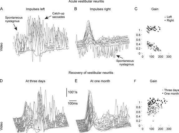
Background: The head impulse test (HIT) is a useful bedside test to identify peripheral vestibular deficits. However, such a deficit of the vestibulo-ocular reflex (VOR) may not be diagnosed because corrective saccades cannot always be detected by simple observation. The scleral search coil technique is the gold standard for HIT measurements, but it is not practical for routine testing or for acute patients, because they are required to wear an uncomfortable contact lens.

Objective: To develop an easy-to-use video HIT system (vHIT) as a clinical tool for identifying peripheral vestibular deficits. To validate the diagnostic accuracy of vHIT by simultaneous measures with video and search coil recordings across healthy subjects and patients with a wide range of previously identified peripheral vestibular deficits.

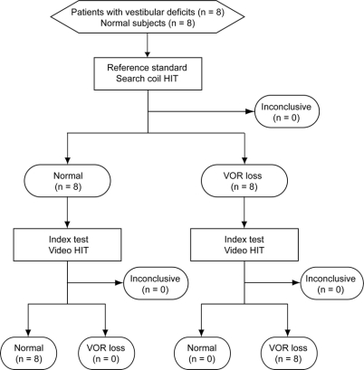
Methods: Horizontal HIT was recorded simultaneously with vHIT (250 Hz) and search coils (1,000 Hz) in 8 normal subjects, 6 patients with vestibular neuritis, 1 patient after unilateral intratympanic gentamicin, and 1 patient with bilateral gentamicin vestibulotoxicity.

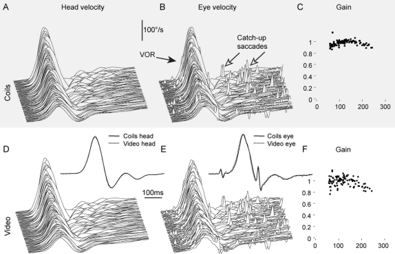
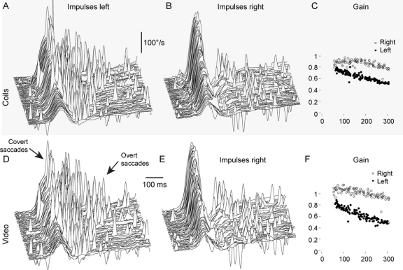
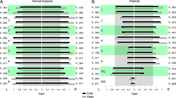
Results: Simultaneous video and search coil recordings of eye movements were closely comparable (average concordance correlation coefficient r(c) = 0.930). Mean VOR gains measured with search coils and video were not significantly different in normal (p = 0.107) and patients (p = 0.073). With these groups, the sensitivity and specificity of both the reference and index test were 1.0 (95% confidence interval 0.69-1.0). vHIT measures detected both overt and covert saccades as accurately as coils.

Conclusions: The video head impulse test is equivalent to search coils in identifying peripheral vestibular deficits but easier to use in clinics, even in patients with acute vestibular neuritis.


Figures

None
Figure 1 Flow chart for the comparison of video and search coil measures of head impulses HIT = head impulse test; VOR = vestibulo-ocular reflex.
None
Figure 2 Simultaneous video and search coil recordings of a horizontal head impulse test in a normal subject Simultaneous angular head velocity recordings of a search coil mounted on a dental impression tray (A) and a gyroscope mounted on the video glasses (D) during graded horizontal head impulses. The close similarity between the 2 recordings demonstrates minimal slippage of the video glasses relative to the head. Simultaneous angular eye velocity recordings of a scleral search coil (B) and high-speed video-oculography (E) of the same eye. Both recording techniques accurately record the vestibulo-ocular reflex (VOR) and detect even the smallest catch-up saccades. Normal VOR gains of individual head impulses are comparable with search coil recording (C) and video-oculography (F). Scleral search coil recording is sampled at 1,000 Hz (A and B), video-oculography is sampled at 250 Hz (D and E), and both are plotted on the same time scale. Head and eye velocity traces from individual impulses are stacked according to increasing peak head velocity. (Insets D and E) Simultaneous video and search coil recordings are shown superimposed to facilitate comparison of single head and eye velocity traces.
None
Figure 3 Simultaneous video and search coil recordings of a horizontal head impulse test in a patient after left vestibular neuritis (A and D) Head impulses to the left (affected) side demonstrate the reduced vestibulo-ocular reflex (VOR) response. Both recording methods detect covert saccades during head rotation and overt saccades after head rotation (arrows). The pattern of catch-up saccades is identical for both recording methods. (B and E) Both recording methods demonstrate an almost normal VOR response to the healthy right side, with small overt saccades after head rotation. (C and F) Both recording methods clearly differentiate the reduced VOR gains of the left affected side (filled circles) from the right healthy side (empty circles). (A, B, D, and E) Signs of eye velocity traces are inverted so that VOR responses and catch-up saccades always point upward.
None
Figure 4 VOR gain measures with search coils compared with video-oculography in normal subjects and patients with peripheral vestibular deficits (A) The vestibulo-ocular reflex (VOR) gain for healthy subjects is almost identical for the 2 different methods of measurement. The 2 sets of data give highly reproducible values of VOR gain. (B) Video-oculography identifies the affected side in vestibular neuritis (1–6) and intratympanic gentamicin (ITG, 7) patients as reliably as search coil measurements. Patient 8 with bilateral vestibular loss (BVL) due to systemic gentamicin vestibulotoxicity demonstrates reproducibility of both methods at very low VOR gains. Bar graphs show the mean VOR gain measures with search coils (black bars) compared with video-oculography (white bars). For each individual, 2 data sets were recorded in the same session. Concordance correlation coefficients (rc) index the similarity between search coil and video-oculography measurements. The vertical gray box indicates deficient VOR gain values (cutoff gain 0.68).
None
Figure 5 Diagnosis of acute vestibular neuritis and documentation of recovery (A–C) Video head impulse test of a patient 2 days after onset of acute vestibular neuritis. (A) In head impulses to the affected left side, catch-up saccades replace the deficient vestibulo-ocular reflex (VOR). The spontaneous nystagmus (scattered spikes) beats in the same direction as the catch-up saccades. (B) In head impulses to the healthy right side, the VOR is preserved and the spontaneous nystagmus beats to the opposite direction. (C) The VOR gain is deficient to the left (open squares) but preserved to the right (filled squares). (D–F) Video head impulse test of a patient 3 days (D) and 1 month (E) after onset of acute vestibular neuritis. Between the 2 recordings, the VOR gains returned toward normal (F), the majority of catch-up saccades disappeared, and the patient recovered from symptoms. (A, B, D, and E) Signs of eye velocity traces are inverted so that VOR responses and catch-up saccades always point upward.

References

    1. Halmagyi GM, Curthoys IS. A clinical sign of canal paresis. Arch Neurol 1988;45:737–739. - PubMed
    1. Aw ST, Fetter M, Cremer PD, Karlberg M, Halmagyi GM. Individual semicircular canal function in superior and inferior vestibular neuritis. Neurology 2001;57:768–774. - PubMed
    1. Schmid-Priscoveanu A, Bohmer A, Obzina H, Straumann D. Caloric and search-coil head-impulse testing in patients after vestibular neuritis. J Assoc Res Otolaryngol 2001;2:72–78. - PMC - PubMed
    1. Jorns-Haderli M, Straumann D, Palla A. Accuracy of the bedside head impulse test in detecting vestibular hypofunction. J Neurol Neurosurg Psychiatry 2007;78:1113–1118. - PMC - PubMed
    1. Black RA, Halmagyi GM, Thurtell MJ, Todd MJ, Curthoys IS. The active head-impulse test in unilateral peripheral vestibulopathy. Arch Neurol 2005;62:290–293. - PubMed