Review
. 2009 Dec 1;587(Pt 23):5591-600.
doi: 10.1113/jphysiol.2009.178350. Epub 2009 Oct 5.

Cell-cell and intracellular lactate shuttles

Affiliations
Review

Cell-cell and intracellular lactate shuttles

George A Brooks. J Physiol. .

Abstract

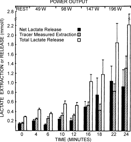
Once thought to be the consequence of oxygen lack in contracting skeletal muscle, the glycolytic product lactate is formed and utilized continuously in diverse cells under fully aerobic conditions. 'Cell-cell' and 'intracellular lactate shuttle' concepts describe the roles of lactate in delivery of oxidative and gluconeogenic substrates as well as in cell signalling. Examples of the cell-cell shuttles include lactate exchanges between between white-glycolytic and red-oxidative fibres within a working muscle bed, and between working skeletal muscle and heart, brain, liver and kidneys. Examples of intracellular lactate shuttles include lactate uptake by mitochondria and pyruvate for lactate exchange in peroxisomes. Lactate for pyruvate exchanges affect cell redox state, and by itself lactate is a ROS generator. In vivo, lactate is a preferred substrate and high blood lactate levels down-regulate the use of glucose and free fatty acids (FFA). As well, lactate binding may affect metabolic regulation, for instance binding to G-protein receptors in adipocytes inhibiting lipolysis, and thus decreasing plasma FFA availability. In vitro lactate accumulation upregulates expression of MCT1 and genes coding for other components of the mitochondrial reticulum in skeletal muscle. The mitochondrial reticulum in muscle and mitochondrial networks in other aerobic tissues function to establish concentration and proton gradients necessary for cells with high mitochondrial densities to oxidize lactate. The presence of lactate shuttles gives rise to the realization that glycolytic and oxidative pathways should be viewed as linked, as opposed to alternative, processes, because lactate, the product of one pathway, is the substrate for the other.


Figures

Figure 1
Figure 1. Lactate disposal (Ri) and oxidation (Rox) rates plotted as functions of oxygen consumption rate () in 6 men at rest and exercise power outputs eliciting 50 and 75% of
Values are means ±s.e.m. Reprinted from Mazzeo et al. (1986) with permission of the American Physiological Society.
Figure 2
Figure 2. Net lactate release, tracer-measured lactate extraction and total lactate release (extraction + net release) in working leg muscles as a function of time
Net lactate release underestimates total intramuscular turnover at all times. Values are means ±s.e.m., n= 6 for all but the last sample when n= 3. Reprinted from Stanley et al. (1986) with permission of the American Physiological Society.
Figure 3
Figure 3. Effects of exercise intensity and training on lactate on lactate metabolic clearance rate (MCR)
Values are means ±s.e.m. for 8–9 subjects. Reprinted from Bergman et al. (1999) with permission of the American Physiological Society.
Figure 4
Figure 4. MCT1 message (A), and protein levels (B) in L6 cells after 1 h incubation at the indicated [lactate] levels
A good correlation between message and protein levels is apparent. Protein levels determined from Western blotting and mRNA from RT-PCR. Data from Hashimoto et al. (2007).
Figure 5
Figure 5. Immunohistochemical images demonstrating some components of the lactate oxidation complex in cultured L6 muscle cells
This complex involves the mitochondrial constituent cytochrome oxidase (COX), the lactate–pyruvate transport protein (MCT1), lactate dehydrogenase (LDH) and other constituents. A, co-localization of MCT1 and the mitochondrial reticulum. MCT1 was detected at both sarcolemmal and intracellular domains (A1). Using MitoTracker the mitochondrial reticulum was extensively elaborated and detected at intracellular domains throughout L6 cells (A2). When signals from probes for the lactate transporter (MCT1, green, A1) and mitochondria (red, A2) were merged, superposition of the signals (yellow) showed co-localization of MCT1 and components of the mitochondrial reticulum, particularly at perinuclear cell domains (A3). B, lactate dehydrogenase (LDH) (B1) and mitochondrial cytochrome oxidase (COX) (B2) are imaged. Superposition of signals for LDH (red, B1) and COX (green, B2) shows co-localization of LDH in the mitochondrial reticulum (yellow) of cultured L6 rat muscle cells (D3). Depth of field ∼1 μm, scale bar = 10 μm. Reprinted from Hashimoto et al. (2006) with permission of the American Physiological Society.
Figure 6
Figure 6. Cellular locations of MCT1 and MCT2 lactate transporter isoforms and the mitochondrial reticulum (cytochrome oxidase, COX) in adult rat plantaris muscle determined using confocal laser scanning microscopy (CLSM) and fluorescent probes for the respective proteins
Comparisons for MCT1 are shown in the first row (A1–A3), and for MCT2 in the second row (B1–B3). The localization of COX was detected in rat plantaris muscle (A1 and B1). MCT1 was detected throughout the cells including subsarcolemmal (arrowheads) and interfibrillar (arrows) domains (A2). MCT1 abundance was greatest in oxidative fibres where COX is abundant and the signal strong. When these MCT1 (green) and COX (red) were merged, superposition of the two probes was clear (yellow), a finding prominent at interfibrillar (arrows) as well as sarcolemmal (arrowheads) cell domains (A3). In contrast, the signal for MCT2 (B2) was weak, relatively more noticeable in fibres denoted by strong staining for COX (B1 and B3, broken line is delineated around oxidative fibre to distinguish the faint signal for MCT2). Overlap of MCT2 and COX is insignificant, denoted by absence of yellow in B3. Scale bar = 50 μm. Sections are from the same animal. Reprinted from Hashimoto et al. (2005).

References

    1. Baba N, Sharma HM. Histochemistry of lactic dehydrogenase in heart and pectoralis muscles of rat. J Cell Biol. 1971;51:621–635. - PMC - PubMed
    1. Bergman BC, Horning MA, Casazza GA, Wolfel EE, Butterfield GE, Brooks GA. Endurance training increases gluconeogenesis during rest and exercise in men. Am J Physiol Endocrinol Metab. 2000;278:E244–251. - PubMed
    1. Bergman BC, Wolfel EE, Butterfield GE, Lopaschuk G, Casazza GA, Horning MA, Brooks GA. Active muscle and whole body lactate kinetics after endurance training in men. J Appl Physiol. 1999;87:1684–1696. - PubMed
    1. Bertocci LA, Lujan BF. Oxidative incorporation and utilization of [3-13C]lactate and [1,2-13C]acetate by rat skeletal muscle. J Appl Physiol. 1999;86:2077–2089. - PubMed
    1. Brandt RB, Laux JE, Spainhour SE, Kline ES. Lactate dehydrogenase in mitochondria. Arch Biochem Biophys. 1987;259:412–422. - PubMed