Clinical Trial
. 2009 Nov;119(11):3226-35.
doi: 10.1172/JCI38374. Epub 2009 Oct 1.

Adoptive immunotherapy with liver allograft-derived lymphocytes induces anti-HCV activity after liver transplantation in humans and humanized mice

Affiliations
Clinical Trial

Adoptive immunotherapy with liver allograft-derived lymphocytes induces anti-HCV activity after liver transplantation in humans and humanized mice

Masahiro Ohira et al. J Clin Invest. 2009 Nov.

Abstract

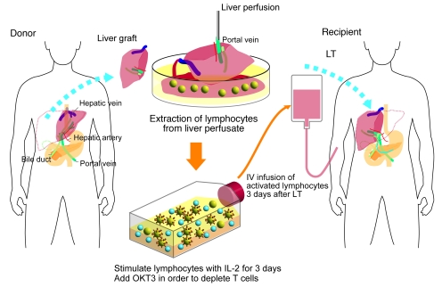
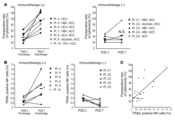
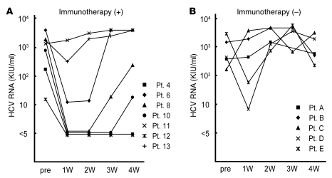
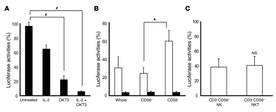
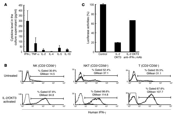
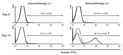
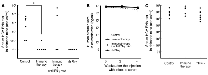
After liver transplantation in HCV-infected patients, the virus load inevitably exceeds pre-transplantation levels. This phenomenon reflects suppression of the host-effector immune responses that control HCV replication by the immunosuppressive drugs used to prevent rejection of the transplanted liver. Here, we describe an adoptive immunotherapy approach, using lymphocytes extracted from liver allograft perfusate (termed herein liver allograft-derived lymphocytes), which includes an abundance of NK/NKT cells that mounted an anti-HCV response in HCV-infected liver transplantation recipients, despite the immunosuppressive environment. This therapy involved intravenously injecting patients 3 days after liver transplantation with liver allograft-derived lymphocytes treated with IL-2 and the CD3-specific mAb OKT3. During the first month after liver transplantation, the HCV RNA titers in the sera of recipients who received immunotherapy were markedly lower than those in the sera of recipients who did not receive immunotherapy. We further explored these observations in human hepatocyte-chimeric mice, in which mouse hepatocytes were replaced by human hepatocytes. These mice unfailingly developed HCV infections after inoculation with HCV-infected human serum. However, injection of human liver-derived lymphocytes treated with IL-2/OKT3 completely prevented HCV infection. Furthermore, an in vitro study using genomic HCV replicon-containing hepatic cells revealed that IFN-gamma-secreting cells played a pivotal role in such anti-HCV responses. Thus, our study presents what we believe to be a novel paradigm for the inhibition of HCV replication in HCV-infected liver transplantation recipients.


Figures

Figure 1
Figure 1. Schematic outline of adoptive immuno­therapy with lymphocytes extracted from liver allograft perfusate.
The therapy involved giving an intravenous injection of IL-2/OKT3–treated liver lymphocytes to LT recipients. The lymphocytes were extracted from the donor liver graft perfusate. After 3 days of culture with IL-2 (100 JRU/ml), the activated liver NK cell–enriched lymphocytes were administered to the LT recipients through venous circulation. OKT3 (1 μg/ml) was added to the culture medium 1 day before this administration in order to prevent GVHD.
Figure 2
Figure 2. Adoptive immunotherapy with IL-2/OKT3–treated liver lymphocytes promoted the cytotoxic activity and TRAIL expression of NK cells in LT recipients.
(A) The NK cytotoxic activities of the indicated effectors against their target cells were analyzed by the 51Cr-release assay. The dot plot represents the NK cytotoxic activities of freshly isolated peripheral blood lymphocytes obtained from recipients who received immunotherapy (+) ( n = 7) and did not receive immunotherapy (–) (n = 5) against HepG2 target cells (effector/target [E/T] ratio, 40:1) 3 and 7 days after LT. NK cytotoxic activities are represented as a proportion (percentage) of the preoperative cytotoxicity in each patient. Horizontal lines indicate the mean. Statistical analyses were performed using the 2-tailed, paired Student’s t test. *P < 0.05 for day 7 versus day 3. (B) The frequency of TRAIL+ NK cells increased remarkably in the peripheral blood of LT recipients who received the immunotherapy. Horizontal lines indicate the mean. Statistical analyses were performed using the Mann-Whitney U test. #P = 0.013 for immunotherapy group versus untreated group in postoperative day 7. (C) Correlation between TRAIL+ NK cell ratio and NK cytolytic activity after LT (Spearman rank-order correlation coefficient = 0.54, P = 0.01). Statistical analyses were performed using the Spearman rank-order correlation coefficient. The diagonal line indicates a linear regression line. Each dot indicates the cytotoxicity and TRAIL+ NK cell percentange of each patient. C1, control 1; POD, postoperative day; Pt., patient.
Figure 3
Figure 3. Serial measurement of the HCV RNA titers of LT recipients after LT.
The HCV RNA titers in the sera of LT recipients who received immunotherapy were markedly lower than those in the sera of LT recipients who did not receive the therapy during the first month after LT. Each line with a different symbol represents serial HCV RNA titers from an LT recipient who received (+) (A; n = 7) and 1 who did not receive (–) (B; n = 5) the immunotherapy after LT. KIU, kilo international unit; pre, pre LT; W, week.
Figure 4
Figure 4. The cultivation of liver lymphocytes with IL-2/OKT3 markedly promoted anti-HCV activity.
(A) Activation by IL-2 and OKT3 significantly promoted the anti-HCV effect of the liver allograft–derived lymphocytes that were cultured in complete medium with and without IL-2 (100 JRU/ml) for 3 days. OKT3 (1 μg/ml) was then added 1 day before coculturing with HCV replicon cells, at the indicated time. The bar graphs indicate the luciferase activities of the cells in each group. Data are presented as mean ± SEM (n = 5). Statistical analyses were performed using the Mann-Whitney U test with Bonferroni correction after the Kruskal-Wallis H test. #P < 0.01 for OKT3 and IL-2/OKT3 treatment versus no treatment. (B) CD56+ fraction, including NK and NKT cells, strongly inhibited HCV replication. The culture conditions are described in A. By magnetic cell sorting, CD56+ and CD56 fractions were isolated from the activated lymphocytes and analyzed for anti-HCV activity. The bar graphs indicate the luciferase activities of the cells in each group (IL-2–treated group, white bars; IL-2 plus OKT3–treated group, black bars). Whole, whole lymphocytes. Data are presented as mean ± SEM (n = 5). Statistical analyses were performed using the Mann-Whitney U test. *P < 0.05 for CD56+ fraction versus CD56 fraction. (C) Anti-HCV effect of NK cells was almost identical to that of NKT cells after IL-2 activation. The liver allograft–derived lymphocytes were cultured in complete medium with IL-2 (100 JRU/ml) for 3 days. By magnetic sorting, CD3CD56+ (NK) and CD3+CD56+ (NKT) fractions were isolated from the activated lymphocytes and analyzed for anti-HCV activity. Data are presented as mean ± SEM (n = 6).
Figure 5
Figure 5. Anti-HCV activity of IL-2/OKT3–treated liver lymphocytes was dependent on their IFN-γ secretion ability.
(A) IFN-γ was the major cytokine released from the cultured cells. The bar graphs indicate the concentrations of various cytokines (IFN-γ, TNF-α, IL-2, IL-4, IL-5, and IL-10) detected in the coculture supernatant by CBA. Data are presented as mean ± SEM (n = 3). (B) The effects of IL-2 and OKT3 (100 JRU/ml and 1 μg/ml, respectively) on IFN-γ production by stimulated CD3CD56+ NK, CD3+CD56+ NKT, and CD3+CD56 T cells were evaluated by a combination of cell surface and cytoplasmic mAb staining and subsequent flow cytometric analysis. Histograms represent the log fluorescence intensities obtained upon staining for IFN-γ after gating of each fraction. Dotted lines represent negative control staining with isotype-matched mAbs. Horizontal lines indicate the gated portion of lymphocytes. GMean, geometric mean fluorescent intensity. (C) Blocking of IFN-γ with mAb (100 μg/ml) elucidated the marked role played by IFN-γ in producing the anti-HCV effect. The bar graphs indicate the luciferase activities of the cells in each group. Data are presented as mean ± SEM of a representative triplicate sample.
Figure 6
Figure 6. Adoptive immunotherapy with IL-2/OKT3–treated liver lymphocytes induced the production of IFN-γ in the LT recipients.
At 14 days after LT, the number of IFN-γ–secreting cells in the peripheral blood of LT recipients treated with the adoptive immunotherapy (+) with IL-2/OKT3–treated liver lymphocytes, including NK and NKT cells, was significantly higher than that in the peripheral blood of untreated LT recipients (–). Histograms represent the proportion (percentage) of IFN-γ–positive cells among the mononuclear cells obtained from the peripheral blood of the immunotherapy (n = 4) and control group (n = 4) LT recipients. Dotted lines represent negative control staining with isotype-matched mAbs. Horizontal lines indicate the gated portion of lymphocytes. Data are presented as mean ± SEM. Histogram profiles shown are representative of 4 independent experiments. Statistical analyses were performed using the Mann-Whitney U test. *P < 0.05 for immunotherapy group versus control group.
Figure 7
Figure 7. Adoptive immunotherapy with IL-2/OKT3–treated liver lymphocytes prevented HCV infection in human hepatocyte–chimeric mice.
(A) Human hepatocyte–chimeric mice were intravenously injected with human serum samples positive for HCV genotype 1b. Two weeks after injecting the infected serum, the mice were intraperitoneally inoculated with IL-2/OKT3–treated liver lymphocytes (20 × 106 cells/mouse; n = 6) for adoptive immunotherapy. When indicated, anti-human IFN-γ mAb was injected intraperitoneally 1 day before the immunotherapy (n = 4). Intraperitoneal injection of recombinant human IFN-γ (rhIFN-γ) was commenced at 2 weeks after injecting the infected serum (n = 5). The untreated mice served as controls (n = 6). The dot plots represent serum HCV RNA titers in each chimeric mouse 4 weeks after the injecting the infected serum. Statistical analyses were performed using the Mann-Whitney U test. *P < 0.01 for immunotherapy group versus control group. (B) The lines represent serial changes in human serum albumin levels in the sera of the mice indicated above. Data are presented as mean ± SEM. (C) IL-2/OKT3–treated liver lymphocytes (20 × 106 cells/mouse) were intraperitoneally inoculated 4 weeks after the injection with the infected serum (n = 5) for adoptive immunotherapy. Intraperitoneal injection of recombinant human IFN-γ was commenced 4 weeks after the injecting the infected serum (n = 9). The untreated mice served as controls (n = 9). The dot plots represent serum HCV RNA titers in each chimeric mouse 6 weeks after injection with the infected serum.

Comment in

Similar articles

Cited by

References

    1. Petrovic L.M. Early recurrence of hepatitis C virus infection after liver transplantation. Liver Transpl. 2006;12:S32–S37. doi: 10.1002/lt.20942. - DOI - PubMed
    1. Brown R.S. Hepatitis C and liver transplantation. Nature. 2005;436:973–978. doi: 10.1038/nature04083. - DOI - PubMed
    1. Garcia-Retortillo M., et al. Hepatitis C virus kinetics during and immediately after liver transplantation. Hepatology. 2002;35:680–687. doi: 10.1053/jhep.2002.31773. - DOI - PubMed
    1. Berenguer M. Natural history of recurrent hepatitis C. Liver Transpl. 2002;8:S14–S18. doi: 10.1053/jlts.2002.35781. - DOI - PubMed
    1. Ishiyama K., et al. Difference in cytotoxicity against hepatocellular carcinoma between liver and periphery natural killer cells in humans. Hepatology. 2006;43:362–372. doi: 10.1002/hep.21035. - DOI - PubMed

Publication types

MeSH terms