. 2009 Oct;7(10):e1000213.
doi: 10.1371/journal.pbio.1000213. Epub 2009 Oct 6.

Rational extension of the ribosome biogenesis pathway using network-guided genetics

Affiliations

Rational extension of the ribosome biogenesis pathway using network-guided genetics

Zhihua Li et al. PLoS Biol. 2009 Oct.

Abstract

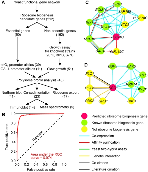
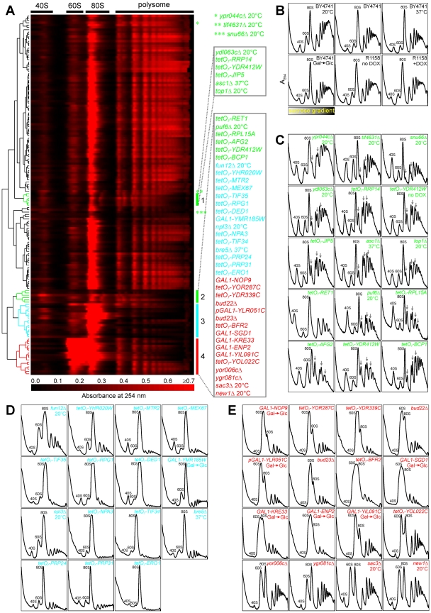
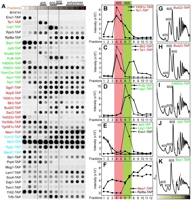
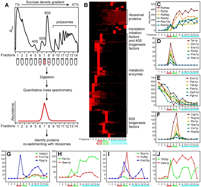
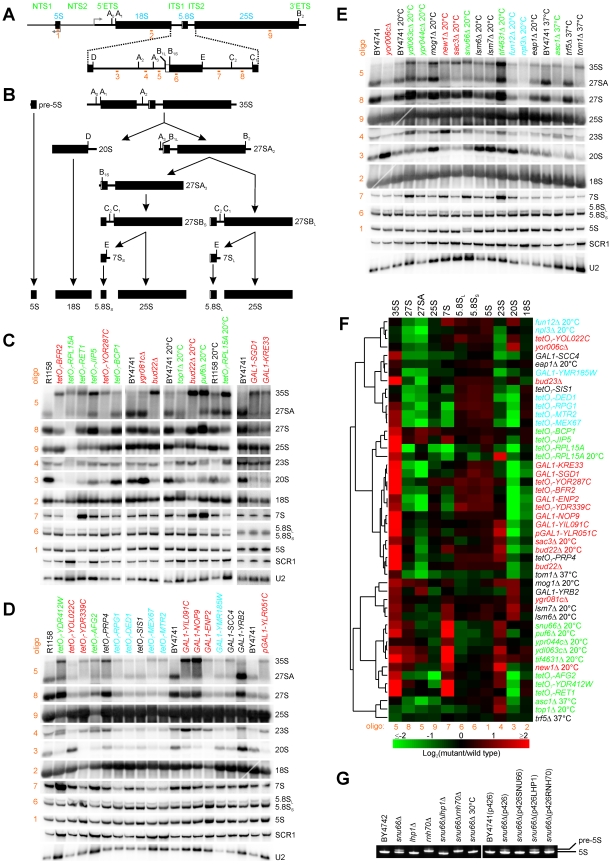
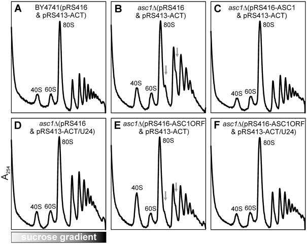
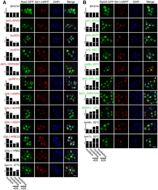
Biogenesis of ribosomes is an essential cellular process conserved across all eukaryotes and is known to require >170 genes for the assembly, modification, and trafficking of ribosome components through multiple cellular compartments. Despite intensive study, this pathway likely involves many additional genes. Here, we employ network-guided genetics-an approach for associating candidate genes with biological processes that capitalizes on recent advances in functional genomic and proteomic studies-to computationally identify additional ribosomal biogenesis genes. We experimentally evaluated >100 candidate yeast genes in a battery of assays, confirming involvement of at least 15 new genes, including previously uncharacterized genes (YDL063C, YIL091C, YOR287C, YOR006C/TSR3, YOL022C/TSR4). We associate the new genes with specific aspects of ribosomal subunit maturation, ribosomal particle association, and ribosomal subunit nuclear export, and we identify genes specifically required for the processing of 5S, 7S, 20S, 27S, and 35S rRNAs. These results reveal new connections between ribosome biogenesis and mRNA splicing and add >10% new genes-most with human orthologs-to the biogenesis pathway, significantly extending our understanding of a universally conserved eukaryotic process.


Conflict of interest statement

The authors have declared that no competing interests exist.

Figures

Figure 1
Figure 1. Overview of the analysis.
(A) A yeast functional-gene network reconstructed from diverse functional genomic and proteomic data was employed to predict genes for ribosome biogenesis. For nonessential genes, growth assays of the deletion mutants under different temperature conditions (20°C, 30°C, and 37°C) were used to identify conditional growth defects, and polysome profiles of these strains were collected under slow-growth conditions. For essential genes, mutants with conditional alleles were subjected to polysome profile analyses after depleting the encoded proteins. Genes affecting the ratio of free 40S to free 60S ribosomal subunits upon deletion of the gene or depletion of the encoded protein were further analyzed by co-sedimentation analyses to assign possible protein association with pre-ribosomal particles, by using Northern blots to assay pre-rRNA processing defects, and by ribosomal subunit export assays. Numbers in parentheses are counts of genes implicated in ribosome biogenesis by each analysis. (B) Assessment of the network-based predictability of ribosome biogenesis genes. The ROC curve (red line) shows cross-validated recovery of known ribosome biogenesis genes based on their network connectivity to one another. True positive ribosome biogenesis genes were manually curated based on Gene Ontology annotation. The network-based prediction is considerably stronger than random expectation (dashed line). (C) and (D) show the top 10 network connections for two predicted ribosome biogenesis genes, YIL091C and SGD1.
Figure 2
Figure 2. Ribosome biogenesis defects were confirmed and classified by polysome profiles of mutant strains.
(A) Hierarchical clustering of mutant polysome profiles (rows), with clusters 1 and 2 representing mutants with 60S subunit biogenesis defects (green), cluster 3 displaying translation defects (cyan), and cluster 4 displaying 40S subunit biogenesis defects (red). Three additional mutants with 60S subunit biogenesis defects are labeled with stars. Each row corresponds to the polysome profile of a single strain, plotting nucleic acid absorbance as a function of position in a sucrose density gradient. Strains were cultured at 30°C unless otherwise indicated. Mutants with tetO7-promoter alleles were cultured in medium with 10 µg/ml doxycycline (+DOX) unless indicated with no DOX. Mutants with GAL1-promoter alleles were first cultured in medium with galactose (Gal) as the carbon source and then diluted into medium with dextrose (Glc) as the carbon source. (B) Polysome profiles of wild-type strains cultured under assayed conditions. BY4741 is the control strain for the nonessential gene-deletion mutants and mutants with GAL1-promoter alleles. R1158 is the control strain for the mutants with tetO7-promoter alleles. Peaks corresponding to 40S and 60S ribosomal subunits and 80S monosomes in the polysome profiles are labeled. (C, D, and E) Polysome profiles of mutants with 60S subunit biogenesis defects, translation defects, and 40S subunit biogenesis defects. Gray arrows indicate halfmer polysome peaks.
Figure 3
Figure 3. Physical association of candidate proteins with ribosomal subunits was measured by co-sedimentation and immunoblot.
(A) Immunoblots of fractions collected from sucrose density gradients for strains carrying TAP-tagged gene alleles. Fractions 4 and 5, 6 and 7, 7 and 8, and 9–12 correspond to the 40S, 60S, 80S, and polysome peaks in the sucrose density gradients, respectively. BY4741 is the negative control, and Tsr1-TAP and Lsg1-TAP are the positive controls for 40S subunit biogenesis factors and 60S subunit biogenesis factors, respectively. Rps3-TAP and Rpl8a-TAP show the locations of small and large ribosomal subunits in the sucrose density gradient, respectively, whereas Eno1-TAP represents the proteins that do not co-sediment with ribosomes. (B–F) show quantitation of the immunoblots for Yil091c-TAP, Bfr2-TAP, Puf6-TAP, Jip5-TAP, and New1-TAP. (G–K) show polysome profile defects for several TAP-tagged strains.
Figure 4
Figure 4. Co-sedimentation of candidate proteins with ribosomal subunits was independently verified using mass spectrometry.
(A) Schematic overview of the experimental design. (B) Hierarchical clustering of abundance profiles of 1,023 proteins (row) identified from fractions (columns) of the sucrose density gradient of wild-type yeast cells. Four distinct clusters are enriched (p<10−8; [83]) for r-proteins, translation-initiation factors and 40S biogenesis factors, metabolic enzymes, and 60S biogenesis factors. Representative profiles are plotted for r-proteins (C), 40S biogenesis factors (D), metabolic enzymes (E), and 60S biogenesis factors (F). (G–J) show profiles for several ribosome biogenesis candidates. Abundance in (C–J) is provided as the frequency of spectral counts (×10,000) of each protein in each fraction; abundance in (B) is further row-normalized.
Figure 5
Figure 5. Characterization of mutant pre-rRNA and rRNA processing defects using quantitative Northern blots.
(A) Oligonucleotide probes (orange numbers; see Table S2 for sequence information) within an rDNA repeat were selected to probe the majority of pre-rRNA and rRNA species generated during the pre-rRNA processing pathway, diagrammed in (B). Precise processing defects associated with each mutant strain were identified by Northern blots (C–E) of different pre-rRNA and rRNA species in wild-type and mutants. Strain label colors are the same as in Figure 2. (F) Global trends among the mutant strains could be observed from hierarchical clustering of mutant strains on the basis of pre-rRNA and rRNA abundances measured from the Northern blots (C–E), with red and green colors representing increased and decreased levels of RNA species, respectively, in mutants relative to corresponding wild-type control strains. The defect of the SNU66Δ strain was examined in more detail in (G). In particular, the temperature-dependent 5S processing defect of this strain could be rescued by deletion of LHP1. 5S rRNA was assayed by 10% TBE-Urea gel and SYBR Gold staining. BY4742 is the wild-type control strain for deletion strains, and BY4741(p426) is the wild-type control strain for over-expression strains. Strains were cultured at 20°C unless otherwise indicated.
Figure 6
Figure 6. The U24 snoRNA is responsible for the 60S biogenesis defect observed in an asc1Δ mutant.
Polysome profile of wild-type strain with two control plasmids was shown in (A). The asc1Δ mutant with two control plasmids showed the 60S biogenesis defect (B), and this defect was recovered by full length intron-containing ASC1 gene (C) or intron snoRNA U24 of ASC1 (D), but not by the coded protein of ASC1, deleted of its intron (E). When the intron snoRNA U24 of ASC1 was put back into the asc1Δ mutant expressing the coded protein of ASC1, the polysome profile recovered to wild-type (F). All strains were cultured at 37°C. pRS416 and pRS413-ACT are the control plasmids; pRS416-ASC1 carries full length ASC1 with both intron and exon; pRS413-ACT/U24 carries the intron sequence of ASC1; and pRS416-ASC1ORF carries the sequence of protein coding region of ASC1. Peaks corresponding to 40S and 60S ribosomal subunits and 80S mono-ribosomes in the polysome profiles were labeled. Gray arrows indicate the halfmer polysomes.
Figure 7
Figure 7. Identification of ribosomal subunit nucleolar and nuclear export defects.
(A) Mutants with observed small subunit-export defects. (B) Mutants with large subunit-export defects. The first row of panel (A) and panel (B) represent the wild-type strain BY4741 cultured at 30°C. Rps2-GFP and Rpl25-GFP are reporters for the ribosomal small and large subunits, respectively. Sik1-mRFP is the reporter for the nucleolus. DAPI was used to stain DNA to visualize the nucleus. The white scale bar represents 5 µm. The normalized enrichment of ribosomal subunits in the nucleus or nucleolus relative to the cytoplasm, calculated relative to the appropriate control strain, is plotted for each strain.
Figure 8
Figure 8. Synthetic ribosome biogenesis defects were observed in a double mutant trf5ΔGAL1-PAP2, suggesting that high-scoring genes not confirmed in the previous experiments may often still participate in ribosome biogenesis.
The trf5Δ mutant was cultured in YPD. GAL1-PAP2 and trf5ΔGAL1-PAP2 were first cultured in YPGal, then diluted into YPD and cultured to early logarithmic phase. Gray arrows indicate halfmer polysomes. Strong 60S biogenesis defects were observed for the double mutants, but not in either single gene mutant.

References

    1. Fatica A, Tollervey D. Making ribosomes. Curr Opin Cell Biol. 2002;14:313–318. - PubMed
    1. Venema J, Tollervey D. Ribosome synthesis in Saccharomyces cerevisiae. Annu Rev Genet. 1999;33:261–311. - PubMed
    1. Tschochner H, Hurt E. Pre-ribosomes on the road from the nucleolus to the cytoplasm. Trends Cell Biol. 2003;13:255–263. - PubMed
    1. Fromont-Racine M, Senger B, Saveanu C, Fasiolo F. Ribosome assembly in eukaryotes. Gene. 2003;313:17–42. - PubMed
    1. Zemp I, Kutay U. Nuclear export and cytoplasmic maturation of ribosomal subunits. FEBS Lett. 2007;581:2783–2793. - PubMed

Publication types

MeSH terms

Substances