. 2009;6(2):85-96.
doi: 10.1900/RDS.2009.6.85. Epub 2009 Aug 10.

Histopathology of type 1 diabetes: old paradigms and new insights

Affiliations

Histopathology of type 1 diabetes: old paradigms and new insights

Ken T Coppieters et al. Rev Diabet Stud. 2009.

Abstract

Although our knowledge on the various aspects of diabetes development in the NOD mouse model is substantial and keeps expanding at a dramatic pace, the dataset on histopathologic features of type 1 diabetes (T1D) in patients remains largely stagnant. Early work has established an array of common aspects that have become epitomic in the absence of new patient material. There is a growing consensus that an updated and more detailed view is required that challenges and expands our understanding. Comprehensive initiatives are currently ongoing to address these issues in pre-diabetic, recent onset and longstanding type 1 diabetic individuals, and some of the old data have been recently revisited. In this review article, we wish to provide an overview of where we stand today and how we can correlate the various cross-sectional studies from the past with contemporary models of the disease. We believe an enhanced understanding of the many histopathological particularities in patients as compared to animal models will ultimately lead, not only to more fundamental insights, but also to an improved ability to translate pre-clinical data from bench to bedside.


Figures

Figure 1
Figure 1. Local early events in islets leading to β-cell death
A: Model for a possible sequence causing the β-cells to undergo apoptosis. From left to right, normal islets are challenged by an environmental factor, presumably viral infection, and upregulate IFN-α. This cytokine exerts its effects in a paracrine fashion, inducing upregulation of MHC Class I in all islet cells and the subsequent influx of β-cell-specific CD8 T cells in genetically at-risk individuals. Finally, β-cells undergo apoptosis and most islets become devoid of β-cells, with potentially only a remnant pool of insulin-deficient β-cells that briefly restores insulin sufficiency during the honeymoon phase. B: Section from the pancreatic head region of a normal 72 year old female individual. Note the abundant quantities of functional β-cells and low levels of MHC Class I+ (HLA-ABC) cells, which are scattered in the exocrine region. C: Section from the pancreatic tail region of a 12 year old male with 1 year of clinical diabetes, who died of ketoacidosis. The strong hyperexpression of MHC Class I (HLA-ABC) on all islet cells is obvious, while many β-cells continue to produce insulin. This islet was found to contain limited numbers of infiltrating CD8 T cells.
Figure 2
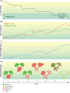
Figure 2. Correlation of histopathology data with the relapsing-remitting hypothesis
A: We described the progression of diabetes development earlier as a stepwise, non-linear decline of β-cell mass over time [53], up to the point where patients lose 60-90% of their functional β-cells and become diabetic. A series of environmental triggers may cause these stages of β-cell decay, explaining why signs of viral infection can be found around the time of diagnosis, following the final step of degeneration. The honeymoon phase is a transient phase of normoglycemia after clinical manifestation, which may serve as a suitable window for therapeutic intervention. B: The stepwise nature of β-cell decay supposedly results from the interplay between auto-reactive T cells and regulatory T cells. Episodes of β-cell loss are caused by a disequilibrium giving auto-reactivity the upper hand. C: Although evidence of significant β-cell proliferations as a rescue mechanism has only been obtained in animal models, the slowly progressing, chronic nature of the prediabetic phase indirectly suggests that some form of β-cell survival takes place. D: Reconciling this model with the combined data from histological reports: 1. One of the earliest histological signs may be the upregulation of MHC Class I in response to an environmental agent. At this point only a minority of islets is affected in a confined pancreatic lobule, and all β-cells remain fully functional. Random sampling such as e.g. by biopsy easily misses out on these subtle events. 2. Some islets become infiltrated by auto-reactive T cells. 3. As regulatory T cells transiently regain supremacy, insulitis subsides leaving a minor fraction of β-cells either death or insulin-deficient. 4. As the prediabetic phase progresses, more islets become MHC Class I+ and insulitis spreads to more lobules. At this point just before clinical onset, the disease should be easily detectable in e.g. pre-screened multiple auto-antibody positive patients. 5. In recent-onset individuals, the majority of β-cell mass is lost, and what is seen histologically are remnants of a process that has been ongoing for years. 6. During the honeymoon phase, some β-cells regain function and support a transient period of normoglycemia. 7. In longstanding cases, the vast majority of islets are devoid of functional β-cells and inflammation, although there is evidence that at least a fraction of patients retain a minor β-cell mass. Together, this stepwise relapsing-remitting course may explain the variability that has been reported between patients at various stages of disease development.

References

    1. Gepts W. Pathologic anatomy of the pancreas in juvenile diabetes mellitus. Diabetes. 1965;14(10):619–633. - PubMed
    1. Bottazzo GF, Florin-Christensen A, Doniach D. Islet-cell antibodies in diabetes mellitus with autoimmune polyendocrine deficiencies. Lancet. 1974;2(7892):1279–1283. - PubMed
    1. MacCuish AC, Irvine WJ, Barnes EW, Duncan LJ. Antibodies to pancreatic islet cells in insulin-dependent diabetics with coexistent autoimmune disease. Lancet. 1974;2(7896):1529–1531. - PubMed
    1. Nerup J, Platz P, Andersen OO, Christy M, Lyngsoe J, Poulsen JE, Ryder LP, Nielsen LS, Thomsen M, Svejgaard A. HL-A antigens and diabetes mellitus. Lancet. 1974;2(7885):864–866. - PubMed
    1. Singal DP, Blajchman MA. Histocompatibility (HL-A) antigens, lymphocytotoxic antibodies and tissue antibodies in patients with diabetes mellitus. Diabetes. 1973;22:429–432. - PubMed

LinkOut - more resources