Review
. 2009:25:457-82.
doi: 10.1146/annurev.cellbio.042308.13350.

Gonad morphogenesis in vertebrates: divergent means to a convergent end

Affiliations
Review

Gonad morphogenesis in vertebrates: divergent means to a convergent end

Tony DeFalco et al. Annu Rev Cell Dev Biol. 2009.

Abstract

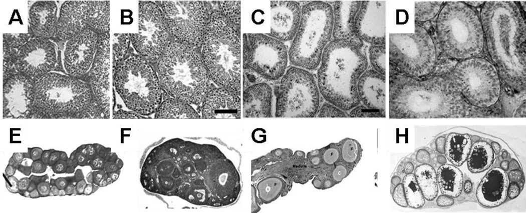
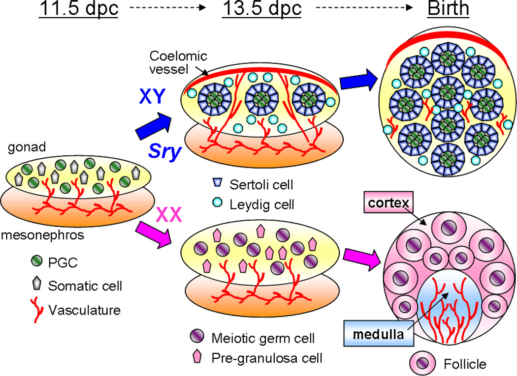
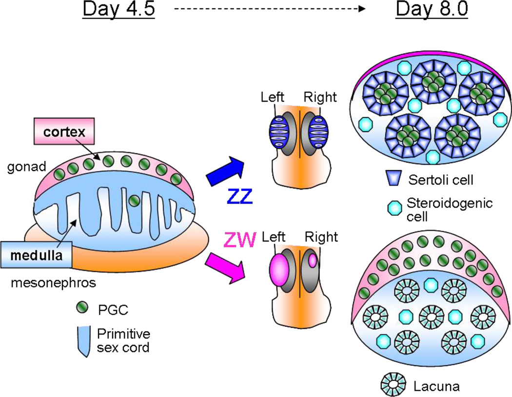
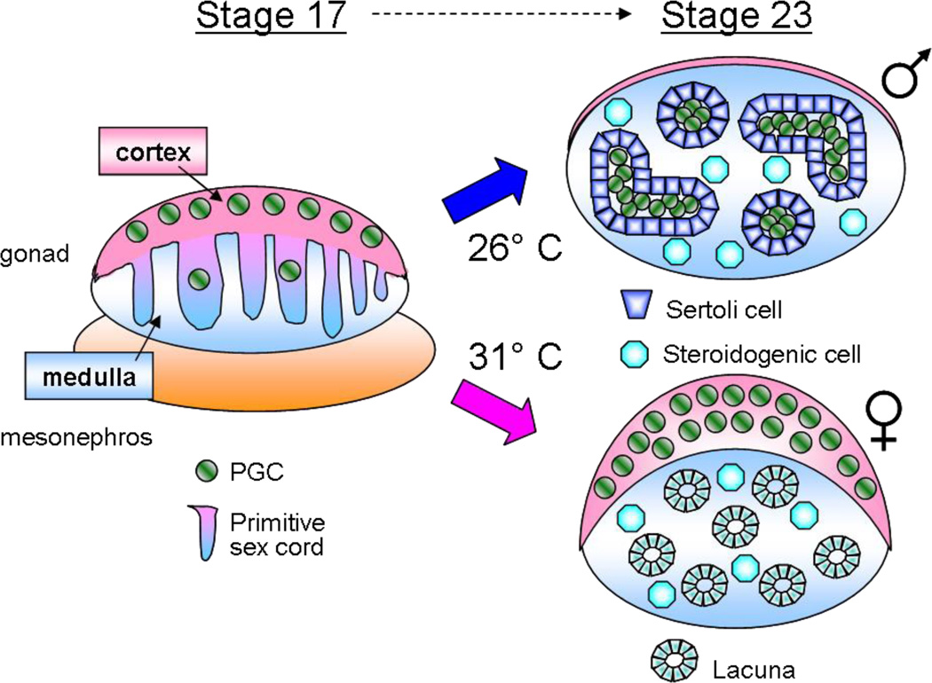
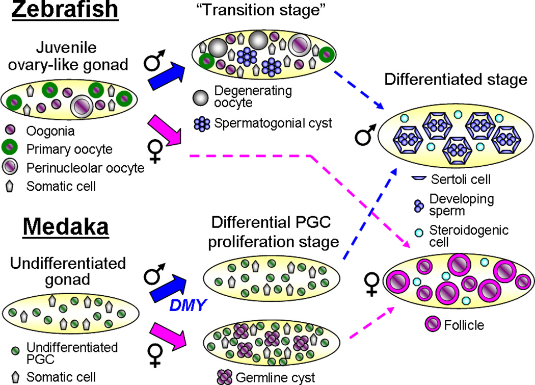
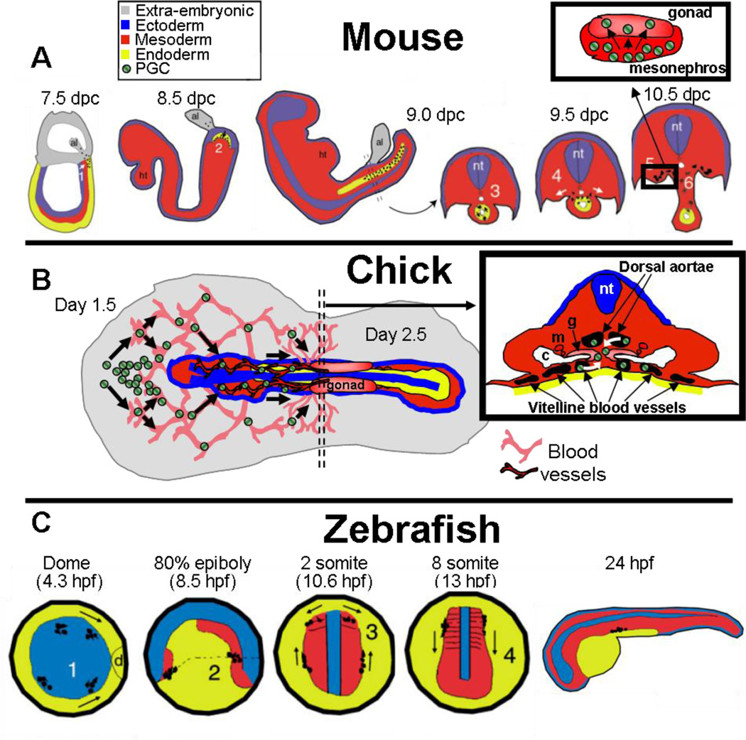
A critical element of successful sexual reproduction is the generation of sexually dimorphic adult reproductive organs, the testis and ovary, which produce functional gametes. Examination of different vertebrate species shows that the adult gonad is remarkably similar in its morphology across different phylogenetic classes. Surprisingly, however, the cellular and molecular programs employed to create similar organs are not evolutionarily conserved. We highlight the mechanisms used by different vertebrate model systems to generate the somatic architecture necessary to support gametogenesis. In addition, we examine the different vertebrate patterns of germ cell migration from their site of origin to colonize the gonad and highlight their roles in sex-specific morphogenesis. We also discuss the plasticity of the adult gonad and consider how different genetic and environmental conditions can induce transitions between testis and ovary morphology.


Figures

Figure 1
Figure 1
Comparison of adult testis (a--d) and ovarian (e--h) structure in various species. (a) Human testis (image courtesy of Dr. Darl Swartz, Department of Animal Science, Purdue University). (b) Mouse testis (image from Zhang et al. 2003, copyright Proceedings of the National Academy of Sciences USA). (c) Chicken testis (image from Song & Silversides 2007, used with permission from Poultry Science). (d) Musk turtle (Sternotherus odoratus) testis (image from Risley 1938, reprinted with permission of Wiley-Liss, Inc., a subsidiary of John Wiley & Sons, Inc.). (e) American leopard frog (Rana pipiens) ovary (image from Hayes et al. 2003, reproduced with permission from Environmental Health Perspectives). (f) Mouse ovary (image from Hosaka et al. 2004, copyright Proceedings of the National Academy of Sciences USA). (g) Chicken ovary (image courtesy of Dr. Thomas Caceci, Virginia-Maryland Regional College of Veterinary Medicine). (h) Medaka ovary (image from Kurokawa et al. 2007, copyright Proceedings of the National Academy of Sciences USA).
Figure 2
Figure 2
Overview of mouse gonad morphogenesis. The expression of Sry directs development of the bipotential gonad toward the testis pathway. Characteristic testis morphology includes formation of testis cords, the coelomic arterial vessel, and Leydig cells. Characteristics of the ovary include entry of germ cells into meiosis, establishment of cortical and medullar domains, and folliculogenesis.
Figure 3
Figure 3
Overview of chick gonad morphogenesis. The bipotential gonad is compartmentalized into a cortex and medulla. Primitive sex cords develop into testis cords in the ZZ (male) gonad and lacunae in the ZW (female) gonad. In the female, only the left gonad grows into a functional ovary. The cortical domain expands in the ovary, whereas it regresses in the testis.
Figure 4
Figure 4
Overview of turtle (T. scripta) gonad morphogenesis. Incubation of eggs at 26°C, the male-producing temperature (MPT), leads to regression of the cortical domain, whereas incubation at 31°C, the female-producing temperature (FPT), leads to expansion of the cortical domain. Primitive sex cords are continuous with the coelomic epithelium and exist in all gonads at the bipotential stage. They become testis cords at the MPT and lacunae at the FPT.
Figure 5
Figure 5
Overview of zebrafish (D. rerio) and medaka (O. latipes) gonad morphogenesis. All undifferentiated zebrafish gonads have ovarian characteristics and undergo a transition in males in which oocytes degenerate while spermatogenic cysts begin to form. The absence of this transition state indicates commitment to ovarian development. In medaka, increased PGC proliferation and generation of cyst-like germ cell clusters are the first indicators of female sex determination and occur prior to any somatic sexual dimorphism. In gonads differentiating as testes, PGC proliferation is much lower.
Figure 6
Figure 6
Different mechanisms of germ cell migration to the gonad in different species. (a) In mice, germ cells are specified at the base of the allantois (al) and enter the gut endoderm (1–2), after which they are passively transported by gut extension, and migrate out the dorsal mesentery to arrive in the gonadal mesoderm (3–6). ht, heart; nt, neural tube. Inset shows migration of the germ cells through the mesonephros into the gonad. (b) In chick, germ cells aggregate in the anterior extraembryonic region, and then enter into the extraembryonic vascular system. Germ cells then extravasate (or pass through the walls of a blood vessel into the surrounding tissue) through the dorsal aortae or through the extraembryonic vitelline vessels into the region near the coelomic epithelium of the presumptive gonad. c, coelom; g, gonad; m, mesonephric ducts/tubules; nt, neural tube. Germ cells not to scale. (c) In zebrafish, germ cells are specified in four clusters during early cleavage stages. They first move dorsally (d) during gastrulation (1), and then collect at the head-trunk mesoderm border (2). After proceeding toward an intermediate target in the lateral plate mesoderm (3), the germ cells migrate posteriorly to the gonad (4). The details of their entry into the gonad are unknown. Images in (a) and (c) were reproduced from Molyneaux & Wylie 2004, with permission of The International Journal of Developmental Biology. Germ cells in low-magnification images in (a) and (c) are represented by small black dots.
Figure 7
Figure 7
Evidence for plasticity of adult gonad phenotypes. (a) Ovotestes of the female European mole (Talpa occidentalis). In the breeding-season ovotestis (left), about 50% of the gonad is dedicated to ovarian and 50% to testicular tissue. A distinct border is evident (yellow dashed line) between the ovarian portion (ov) with developing follicles and the testicular portion (t) with Leydig cells and testis cords. The nonbreeding-season ovotestis (right) shows a dramatic increase in the size of the testicular portion and a decrease in the ovarian portion, which contains only a few immature follicles (inset, arrows). Images courtesy of Dr. Rafael Jimenez, Department of Genetics, University of Granada, Spain. (b) An adult coral goby (Gobiodon erythrospilus) male control gonad (left) contains seminiferous cords with spermatocytes (sc). When treated with estradiol (right), the male changes sex to female, and the gonad converts to a functional ovary, with previtellogenic oocytes (pv) visible. Images from Kroon et al. 2005. Reprinted with permission of The Royal Society. (c) Wild-type mouse follicle (left) containing an oocyte surrounded by granulosa cells. In an estrogen receptor (ER) α/β knockout mutant mouse (right), folliculogenesis is disrupted and granulosa cells transdifferentiate into Sertoli-like cells that cluster to form testis-like tubules. Images from Couse et al. 1999. Reprinted with permission from the American Association for the Advancement of Science.

References

    1. Albrecht KH, Eicher EM. Evidence that Sry is expressed in pre-Sertoli cells and Sertoli and granulosa cells have a common precursor. Dev. Biol. 2001;240:92–107. - PubMed
    1. Barske LA, Capel B. Blurring the edges in vertebrate sex determination. Curr. Opin. Genet. Dev. 2008;18:499–505. - PMC - PubMed
    1. Bauchinger U, Van’t Hof T, Biebach H. Testicular development during long-distance spring migration. Horm. Behav. 2007;51:295–305. - PubMed
    1. Bauchinger U, Wohlmann A, Biebach H. Flexible remodeling of organ size during spring migration of the garden warbler (Sylvia borin) Zoology (Jena) 2005;108:97–106. - PubMed
    1. Bowles J, Knight D, Smith C, Wilhelm D, Richman J, et al. Retinoid signaling determines germ cell fate in mice. Science. 2006;312:596–600. - PubMed

Publication types

LinkOut - more resources