. 2009 Oct 6:10:126.
doi: 10.1186/1471-2202-10-126.

Therapeutic targets and limits of minocycline neuroprotection in experimental ischemic stroke

Affiliations

Therapeutic targets and limits of minocycline neuroprotection in experimental ischemic stroke

Noriyuki Matsukawa et al. BMC Neurosci. .

Abstract

Background: Minocycline, a second-generation tetracycline with anti-inflammatory and anti-apoptotic properties, has been shown to promote therapeutic benefits in experimental stroke. However, equally compelling evidence demonstrates that the drug exerts variable and even detrimental effects in many neurological disease models. Assessment of the mechanism underlying minocycline neuroprotection should clarify the drug's clinical value in acute stroke setting.

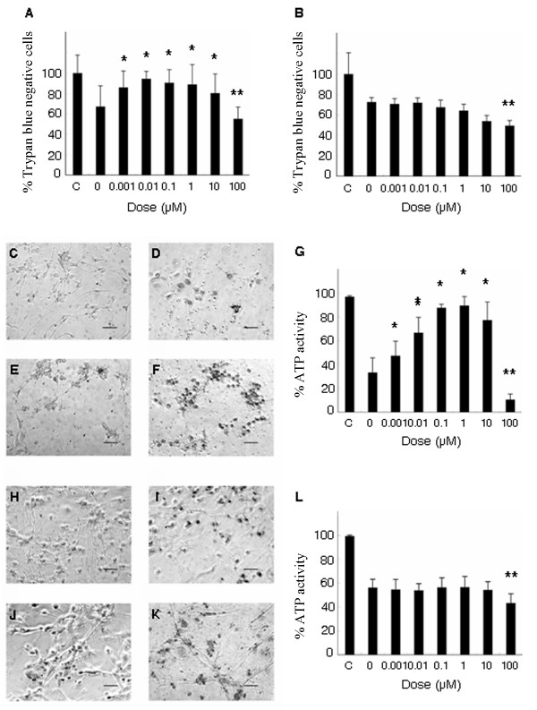
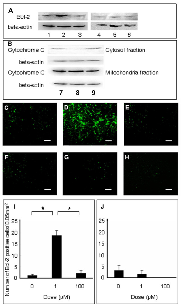
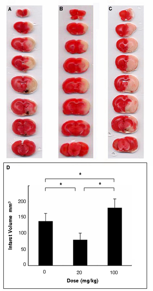
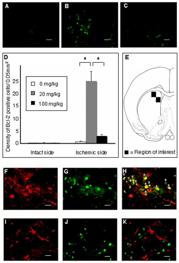
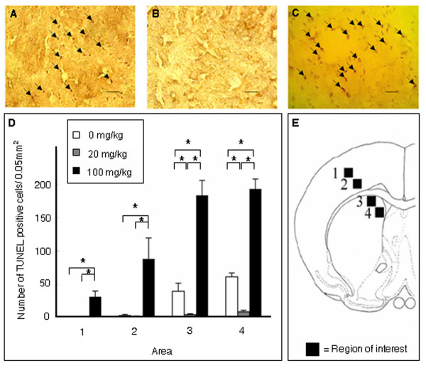
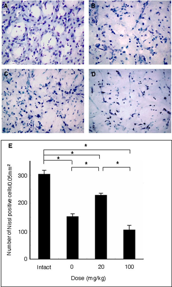
Results: Here, we demonstrate that minocycline attenuates both in vitro (oxygen glucose deprivation) and in vivo (middle cerebral artery occlusion) experimentally induced ischemic deficits by direct inhibition of apoptotic-like neuronal cell death involving the anti-apoptotic Bcl-2/cytochrome c pathway. Such anti-apoptotic effect of minocycline is seen in neurons, but not apparent in astrocytes. Our data further indicate that the neuroprotection is dose-dependent, in that only low dose minocycline inhibits neuronal cell death cascades at the acute stroke phase, whereas the high dose exacerbates the ischemic injury.

Conclusion: The present study advises our community to proceed with caution to use the minimally invasive intravenous delivery of low dose minocycline in order to afford neuroprotection that is safe for stroke.


Figures

Figure 1
Figure 1
ATP activity and trypan blue assay in cultured neurons and astrocytes. ATP activity was measured to reveal cell viability with varying concentrations of minocycline or vehicle. OGD condition reduced ATP activity of cultured neurons to 40% of non-OGD control group (A). Low doses of minocycline (0.001 to 10 μM) preserved cell viability of neurons (A), but not astrocytes (B). In contrast, high dose of minocycline (100 μM) displayed toxicity to both neurons and astrocytes (A, B). In parallel with ATP assay, Trypan blue assay revealed that low doses of minocycline (0.001-10 μM) exerted neuroprotective effects on cultured neurons, but not astrocytes (C-G: neurons, H-L: astrocytes, C, H: non-OGD control, D, I: 0 μM, E, J: 1 μM, F, K: 100 μM of minocycline). High dose of minocycline (100 μM) displayed toxicity for both neurons and astrocytes. Data are shown as mean values ± SEM (*p < 0.05 increase and **p < 0.05 decrease vs. vehicle-treated cultured neurons or astrocytes; A, G: neurons, B, L: astrocytes). Scale bar: 25 μm.
Figure 2
Figure 2
Caspase3/7 activity and tunel staining in cultured neurons and astrocytes. Low dose minocycline (1 μM) reduced caspase3/7 activity of neurons (A), but not of astrocytes (B). In contrast, high dose minocycline (100 μM) displayed no suppressive effects on caspase 3/7 activity of neurons and increased that of astrocytes (A, B). Similarly, low dose minocycline reduced, whereas high dose minocycline increased the number of TUNEL positive neurons compared to vehicle-treated cultured neurons (OGD-exposed neurons treated with 0, 1 and 100 μM of minocycline; panels C, D and E, respectively). On the other hand, minocycline at all doses did not reduce the number of TUNEL positive astrocytes (OGD-exposed astrocytes treated with 0, 1 and 100 μM of minocycline; panels F, G and H, respectively). Data are shown as mean values ± SEM (*p < 0.05. A, I: neurons, B, J: astrocytes). Scale bar: 50 μm.
Figure 3
Figure 3
Bcl-2 expression and cytochrome c release in cultured neurons and astrocytes. Western blotting revealed that low dose minocycline upregulated Bcl-2 expression of neurons (A: lysates from 0, 1 and 100 μM of minocycline-treated neurons correspond to lanes 1, 2, and 3, respectively) with subsequent inhibition cytochrome c release from mitochondria to cytosol (B: lysates from 0, 1 and 100 μM of minocycline-treated neurons correspond to lanes 7, 8, and 9, respectively). In contrast, minocycline at all doses did not upregulate Bcl-2 expression in astrocytes (A: lysates from 0, 1 and 100 μM of minocycline-treated astrocytes correspond to lanes 4, 5, and 6, respectively). Immunocytochemical analysis revealed that minocycline low dose (D: 1 μM) significantly increased the number of Bcl-2 positive neurons compared to vehicle-treated (C: 0 μM) or high dose-treated neurons (E: 100 μM). In contrast, minocycline at all doses did not alter the number of Bcl-2 positive astrocytes (F: 0 μM, G: 1 μM and H: 100 μM). Quantitative analyses of Bcl-2 positive cells are shown in panels I and J. Data represent mean values ± SEM (* p < 0.05. I: neurons, J: astrocytes). Scale bar: 25 μm.
Figure 4
Figure 4
Motor and neurological performance of stroke rats. Both motor and neurological dysfunctions were significantly ameliorated by low dose minocycline (20 mg/kg, i.v.), as revealed by elevated body swing test (EBST; A) and Bederson test (B). In contrast, high dose minocycline (100 mg/kg, i.v.) significantly exacerbated neurological deficits and slightly worsened motor deficits. Data are shown as the mean values ± SEM (*p < 0.05).
Figure 5
Figure 5
Cerebral infarct analysis of stroke brains. TTC staining revealed that low dose minocycline (20 mg/kg; B) significantly reduced cerebral infarct volumes compared to the vehicle group (A). In particular, the striatal infarct volumes in the low dose group were significantly smaller than the vehicle group. In contrast, high dose minocycline (100 mg/kg; C) significantly increased the infarct volumes compared to those in the vehicle group. Quantitative analyses are shown in panel D. Data represent mean values ± SEM (*p < 0.05).
Figure 6
Figure 6
Tunel staining in the ischemic peri-infarct area. Low dose significantly decreased (B), whereas high dose significantly increased (C) the number of TUNEL positive cells in the ischemic striatal peri-infarct area of minocycline-treated stroke rats compared to the vehicle-treated stroke rats (A). Quantitative analyses of Bcl-2 positive cells are shown in panel D. Data are shown as mean values ± SEM (*p < 0.05). Four representative ischemic striatal peri-infarct areas (+0.2 mm anterior to the bregma), in which TUNEL positive cells were counted (data in panel D), are shown in panel E (square boxes labeled 1-4 correspond to areas 1-4 in panel D). Scale bar: 25 μm.
Figure 7
Figure 7
Bcl-2 expression in the ischemic peri-infarct area. Low dose (B) significantly upregulated, whereas high dose (C) significantly suppressed the Bcl-2 expression in the ischemic peri-infarct area of minocycline-treated rats compared to that in the vehicle-treated rats (A). Quantitative analyses of Bcl-2 positive cells are shown in panel D. Data are shown as mean values ± SEM (*p < 0.05). Two representative ischemic striatal peri-infarct areas (+0.2 mm anterior to the bregma), in which Bcl-2 positive cells were counted (data in panel D), are shown in panel E (square boxes). Co-localization of Bcl-2 and MAP2 was found in ischemic striatal peri-infarct area, suggesting anti-apoptotic effects of minocycline via Bcl-2 upregulation in ischemic neurons (F-H). In contrast, GFAP positive astrocytes did not express Bcl-2 (I-K). Scale bar: 25 μm (A-C), 12.5 μm (F-K); asterisks (*): merged cell; green and red immunofluorescent markers correspond to Bcl-2 and MAP2, respectively.
Figure 8
Figure 8
Neuronal survival in the ischemic peri-infarct area. Neuronal survival in the ischemic striatal peri-infarct area, visualized with cresyl violet stain, was significantly preserved by low dose minocycline (A: intact control, B: vehicle, C: 20 mg/kg, D: 100 mg/kg). In contrast, high dose minocycline resulted in the collapse of fundamental neuroarchitecture of the striatum accompanied by severe edema. Data are shown as mean values ± SEM (*p < 0.05). Scale bar: 25 μm.

Similar articles

Cited by

References

    1. Fiskum G, Murphy AN, Beal MF. Mitochondria in neurodegeneration: acute ischemia and chronic neurodegenerative diseases. J Cereb Blood Flow Metab. 1999;19:351–369. doi: 10.1097/00004647-199904000-00001. - DOI - PubMed
    1. Zipfel GJ, Lee JM, Choi DW. Reducing calcium overload in the ischemic brain. N Engl J Med. 1999;341:1543–1544. doi: 10.1056/NEJM199911113412011. - DOI - PubMed
    1. Paschen W. Role of calcium in neuronal cell injury: which subcellular compartment is involved? Brain Res Bull. 2000;53:409–413. doi: 10.1016/S0361-9230(00)00369-5. - DOI - PubMed
    1. Yuan J, Yankner BA. Apoptosis in the nervous system. Nature. 2000;407:802–809. doi: 10.1038/35037739. - DOI - PubMed
    1. Chan PH. Reactive oxygen radicals in signaling and damage in the ischemic brain. J Cereb Blood Flow Metab. 2001;21:2–14. doi: 10.1097/00004647-200101000-00002. - DOI - PubMed

Publication types

MeSH terms