Atrial fibrillation begets atrial fibrillation: autonomic mechanism for atrial electrical remodeling induced by short-term rapid atrial pacing
- PMID: 19808412
- PMCID: PMC2766842
- DOI: 10.1161/CIRCEP.108.784272
Atrial fibrillation begets atrial fibrillation: autonomic mechanism for atrial electrical remodeling induced by short-term rapid atrial pacing
Abstract
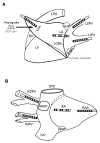
Background: The mechanism(s) for acute changes in electrophysiological properties of the atria during rapid pacing induced atrial fibrillation (AF) is not completely understood. We sought to evaluate the contribution of the intrinsic cardiac autonomic nervous system in acute atrial electrical remodeling and AF induced by 6-hour rapid atrial pacing.
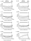
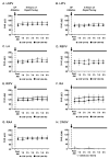
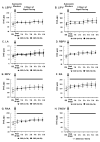
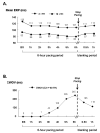
Methods and results: Continuous rapid pacing (1200 bpm, 2x threshold [TH]) was performed at the left atrial appendage. Group 1 (n=7) underwent 6-hour pacing immediately followed by ganglionated plexi (GP) ablation; group 2 (n=7) underwent GP ablation immediately followed by 6-hour pacing; and group 3 (n=4) underwent administration of autonomic blockers, atropine (1 mg/kg), and propranolol (0.6 mg/kg) immediately followed by 6-hour pacing. The effective refractory period (ERP) and window of vulnerability (WOV, in milliseconds), ie, the difference between the longest and the shortest coupling interval of the premature stimulus that induced AF, were measured at 2xTH and 10xTH at the left atrium, right atrium, and pulmonary veins every hour before and after GP ablation or autonomic blockade. In group 1, ERP was markedly shortened in the first 2 hours and then stabilized both at 2xTH and 10xTH; however, WOV was progressively widened throughout the 6-hour period. After GP ablation, ERP was significantly longer than before ablation and AF could not be induced (WOV=0) at either 2xTH or 10xTH. In groups 2 and 3, rapid atrial pacing failed to shorten the ERP. AF could not be induced in 6 of 7 dogs in group 2 and all 4 dogs in group 3 during the 6-hour pacing period.
Conclusions: The intrinsic cardiac autonomic nervous system plays a crucial role in the acute stages of atrial electrical remodeling induced by rapid atrial pacing.
Figures

Similar articles
-
Interactions between atrial electrical remodeling and autonomic remodeling: how to break the vicious cycle.Heart Rhythm. 2012 May;9(5):804-9. doi: 10.1016/j.hrthm.2011.12.023. Epub 2011 Dec 31. Heart Rhythm. 2012. PMID: 22214613
-
Ganglionated plexi modulate extrinsic cardiac autonomic nerve input: effects on sinus rate, atrioventricular conduction, refractoriness, and inducibility of atrial fibrillation.J Am Coll Cardiol. 2007 Jul 3;50(1):61-8. doi: 10.1016/j.jacc.2007.02.066. Epub 2007 Jun 18. J Am Coll Cardiol. 2007. PMID: 17601547
-
The role of ganglionated plexi in apnea-related atrial fibrillation.J Am Coll Cardiol. 2009 Nov 24;54(22):2075-83. doi: 10.1016/j.jacc.2009.09.014. J Am Coll Cardiol. 2009. PMID: 19926016
-
The cardiac autonomic nervous system: A target for modulation of atrial fibrillation.Clin Cardiol. 2019 Jun;42(6):644-652. doi: 10.1002/clc.23190. Epub 2019 May 6. Clin Cardiol. 2019. PMID: 31038759 Free PMC article. Review.
-
Ganglionated plexi as neuromodulation targets for atrial fibrillation.J Cardiovasc Electrophysiol. 2017 Dec;28(12):1485-1491. doi: 10.1111/jce.13319. Epub 2017 Sep 8. J Cardiovasc Electrophysiol. 2017. PMID: 28833764 Free PMC article. Review.
Cited by
-
Effect of renal sympathetic denervation on the inducibility of atrial fibrillation during rapid atrial pacing.J Interv Card Electrophysiol. 2012 Nov;35(2):119-25. doi: 10.1007/s10840-012-9717-y. Epub 2012 Aug 7. J Interv Card Electrophysiol. 2012. PMID: 22869391
-
The immediate trends in atrial electrical remodeling for paroxysmal atrial fibrillation across different modes of catheter ablation.Clin Cardiol. 2021 Jul;44(7):938-945. doi: 10.1002/clc.23617. Epub 2021 Jun 1. Clin Cardiol. 2021. PMID: 34061373 Free PMC article.
-
The effects of atrial ganglionated plexi stimulation on ventricular electrophysiology in a normal canine heart.J Interv Card Electrophysiol. 2013 Jun;37(1):1-8. doi: 10.1007/s10840-012-9774-2. Epub 2013 Feb 8. J Interv Card Electrophysiol. 2013. PMID: 23392954
-
Inflammatory proteomics profiling for prediction of incident atrial fibrillation.Heart. 2023 Jun 14;109(13):1000-1006. doi: 10.1136/heartjnl-2022-321959. Heart. 2023. PMID: 36801832 Free PMC article.
-
An arrhythmogenic metabolite in atrial fibrillation.J Transl Med. 2023 Aug 24;21(1):566. doi: 10.1186/s12967-023-04420-z. J Transl Med. 2023. PMID: 37620858 Free PMC article.
References
-
- Wijffels MC, Kirchhof CJ, Dorland R, Allessie MA. Atrial fibrillation begets atrial fibrillation. A study in awake chronically instrumented goats. Circulation. 1995;92:1954–68. - PubMed
-
- Yue L, Melnyk P, Gaspo R, Wang Z, Nattel S. Molecular mechanisms underlying ionic remodeling in a dog model of atrial fibrillation. Circ Res. 1999;84:776–84. - PubMed
-
- Bosch RF, Scherer CR, Rüb N, Wöhrl S, Steinmeyer K, Haase H, Busch AE, Seipel L, Kühlkamp V. Molecular mechanisms of early electrical remodeling: transcriptional downregulation of ion channel subunits reduces I(Ca,L) and I(to) in rapid atrial pacing in rabbits. J Am Coll Cardiol. 2003;41:858–69. - PubMed
-
- Po SS, Scherlag BJ, Yamanashi WS, Edwards J, Zhou J, Wu R, Geng N, Lazzara R, Jackman WM. Experimental model for paroxysmal atrial fibrillation arising at the pulmonary vein-atrial junctions. Heart Rhythm. 2006;3:201–8. - PubMed
-
- Zhou J, Scherlag BJ, Edwards J, Jackman WM, Lazzara R, Po SS. Gradients of Atrial Refractoriness and Inducibility of Atrial Fibrillation due to Stimulation of Ganglionated Plexi. J Cardiovasc Electrophysiol. 2007;18:83–90. - PubMed
Publication types
MeSH terms
Grants and funding
LinkOut - more resources
Full Text Sources
Other Literature Sources
Medical

