

P wave indices: current status and future directions in epidemiology, clinical, and research applications
- PMID: 19808445
- PMCID: PMC2760837
- DOI: 10.1161/CIRCEP.108.806828
P wave indices: current status and future directions in epidemiology, clinical, and research applications
Abstract
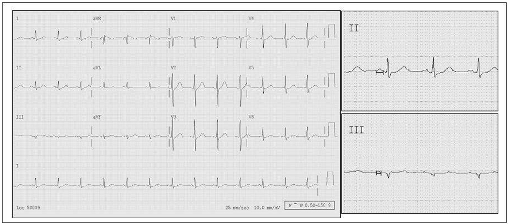
Indices of P wave duration and dispersion are accessible from the surface electrocardiogram. Their prolongation reflects inhomogeneous atrial depolarization secondary to insults such as chronically elevated atrial pressure, ischemia, or metabolic stress. In turn, these insults promote atrial structural remodeling and provide a substrate for atrial fibrillation (AF). P wave indices have been examined in cardiac and non-cardiac disease states. Prolonged P wave indices have been associated with hypertension, obesity and diabetes, all of which are risk factors for AF. Similarly, prolonged P wave duration and dispersion have been associated with AF recurrence in patients with paroxysmal AF and following cardioversion, and with incident AF following cardiothoracic surgeries.
Our review describes the current field of P wave indices. We report the methodology for determining P wave indices. We also describe the strengths and limitations of the current literature on the clinical correlates and prognosis of P wave indices. We suggest future clinical and research directions for P wave indices.
Figures
References
-
- Michelucci A, Bagliani G, Colella A, Pieragnoli P, Porciani MC, Gensini G, Padeletti L. P wave assessment: state of the art update. Card Electrophysiol Rev. 2002;6:215–220. - PubMed
-
- Spach MS. Mounting evidence that fibrosis generates a major mechanism for atrial fibrillation. Circ Res. 2007;101:743–745. - PubMed
-
- Uhley H. It is time to include P-wave duration. Pacing Clin Electrophysiol. 2007;30:293–294. - PubMed
-
- Agarwal YK, Aronow WS, Levy JA, Spodick DH. Association of interatrial block with development of atrial fibrillation. Am J Cardiol. 2003;91:882. - PubMed
-
- Duru M, Seyfeli E, Kuvandik G, Kaya H, Yalcin F. Effect of weight loss on P wave dispersion in obese subjects. Obesity (Silver Spring) 2006;14:1378–1382. - PubMed
Publication types
MeSH terms
Grants and funding
LinkOut - more resources
Full Text Sources
Medical
