

Adenosine stress 64- and 256-row detector computed tomography angiography and perfusion imaging: a pilot study evaluating the transmural extent of perfusion abnormalities to predict atherosclerosis causing myocardial ischemia
- PMID: 19808590
- PMCID: PMC3035629
- DOI: 10.1161/CIRCIMAGING.108.813766
Adenosine stress 64- and 256-row detector computed tomography angiography and perfusion imaging: a pilot study evaluating the transmural extent of perfusion abnormalities to predict atherosclerosis causing myocardial ischemia
Abstract
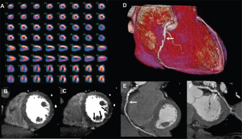
Background: Multidetector computed tomography coronary angiography (CTA) is a robust method for the noninvasive diagnosis of coronary artery disease. However, in its current form, CTA is limited in its prediction of myocardial ischemia. The purpose of this study was to test whether adenosine stress computed tomography myocardial perfusion imaging (CTP), when added to CTA, can predict perfusion abnormalities caused by obstructive atherosclerosis.
Methods and results: Forty patients with a history of abnormal single-photon emission computed tomography myocardial perfusion imaging (SPECT-MPI) underwent adenosine stress 64-row (n=24) or 256-row (n=16) detector CTP and CTA. A subset of 27 patients had invasive angiography available for quantitative coronary angiography. CTA and quantitative coronary angiography were evaluated for stenoses > or =50%, and SPECT-MPI was evaluated for fixed and reversible perfusion deficits using a 17-segment model. CTP images were analyzed for the transmural differences in perfusion using the transmural perfusion ratio (subendocardial attenuation density/subepicardial attenuation density). The sensitivity, specificity, positive predictive value, and negative predictive value for the combination of CTA and CTP to detect obstructive atherosclerosis causing perfusion abnormalities using the combination of quantitative coronary angiography and SPECT as the gold standard was 86%, 92%, 92%, and 85% in the per-patient analysis and 79%, 91%, 75%, and 92% in the per vessel/territory analysis, respectively.
Conclusions: The combination of CTA and CTP can detect atherosclerosis causing perfusion abnormalities when compared with the combination of quantitative coronary angiography and SPECT.
Figures


Comment in
-
Anatomic and functional assessment of coronary artery disease: convergence of 2 aims in a single setting.Circ Cardiovasc Imaging. 2009 May;2(3):163-5. doi: 10.1161/CIRCIMAGING.109.873489. Circ Cardiovasc Imaging. 2009. PMID: 19808586 No abstract available.
References
-
- Miller JM, Rochitte CE, Dewey M, Arbab-Zadeh A, Niinuma H, Gottlieb I, Paul N, Clouse ME, Shapiro EP, Hoe J, Lardo AC, Bush DE, de Roos A, Cox C, Brinker J, Lima JA. Diagnostic performance of coronary angiography by 64-row CT. N Engl J Med. 2008;359:2324–2336. - PubMed
-
- Budoff MJ, Dowe D, Jollis JG, Gitter M, Sutherland J, Halamert E, Scherer M, Bellinger R, Martin A, Benton R, Delago A, Min JK. Diagnostic performance of 64-multidetector row coronary computed tomographic angiography for evaluation of coronary artery stenosis in individuals without known coronary artery disease: results from the prospective multicenter ACCURACY (Assessment by Coronary Computed Tomographic Angiography of Individuals Undergoing Invasive Coronary Angiography) trial. J Am Coll Cardiol. 2008;52:1724–1732. - PubMed
-
- Vanhoenacker PK, Heijenbrok-Kal MH, Van Heste R, Decramer I, Van Hoe LR, Wijns W, Hunink MG. Diagnostic performance of multidetector CT angiography for assessment of coronary artery disease: meta-analysis. Radiology. 2007;244:419–428. - PubMed
-
- Rispler S, Keidar Z, Ghersin E, Roguin A, Soil A, Dragu R, Litmanovich D, Frenkel A, Aronson D, Engel A, Beyar R, Israel O. Integrated single-photon emission computed tomography and computed tomography coronary angiography for the assessment of hemodynamically significant coronary artery lesions. J Am Coll Cardiol. 2007;49:1059–1067. - PubMed
-
- Schuijf JD, Wijns W, Jukema JW, Atsma DE, de Roos A, Lamb HJ, Stokkel MP, Dibbets-Schneider P, Decramer I, De Bondt P, van der Wall EE, Vanhoenacker PK, Bax JJ. Relationship between noninvasive coronary angiography with multi-slice computed tomography and myocardial perfusion imaging. J Am Coll Cardiol. 2006;48:2508–2514. - PubMed
Publication types
MeSH terms
Substances
Grants and funding
LinkOut - more resources
Full Text Sources
Other Literature Sources
Medical
