Multicenter Study
. 2009 May;2(3):191-8.
doi: 10.1161/CIRCIMAGING.108.819938. Epub 2009 Mar 26.

Age-related left ventricular remodeling and associated risk for cardiovascular outcomes: the Multi-Ethnic Study of Atherosclerosis

Affiliations
Multicenter Study

Age-related left ventricular remodeling and associated risk for cardiovascular outcomes: the Multi-Ethnic Study of Atherosclerosis

Susan Cheng et al. Circ Cardiovasc Imaging. 2009 May.

Abstract

Background: Age-related alterations of left ventricular (LV) structure and function that may predispose to cardiovascular events are not well understood.

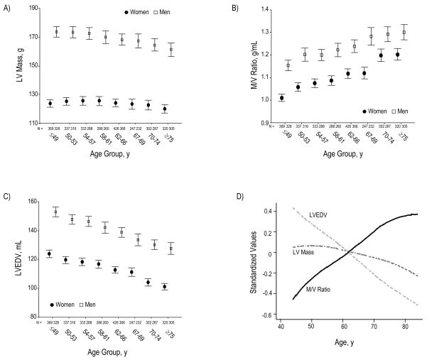
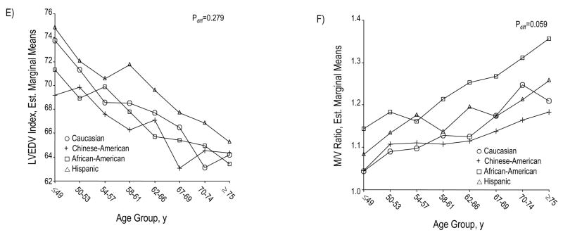
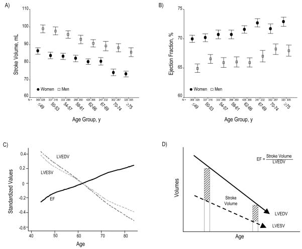
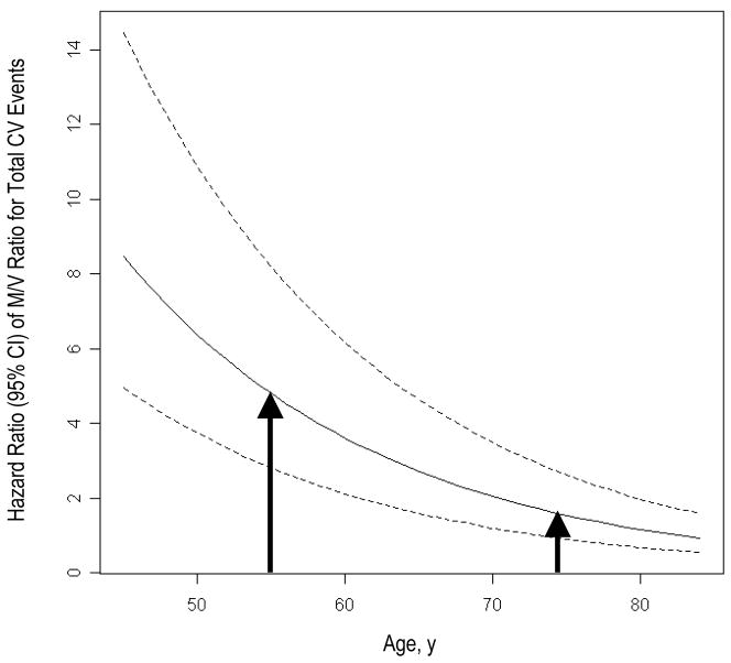
Methods and results: We used cardiac MRI to examine age-related differences in LV structure and function in 5004 participants without overt cardiovascular disease when enrolled in the Multi-Ethnic Study of Atherosclerosis; 1099 participants received additional strain analyses by MRI tagging. We also assessed the relation of age-associated remodeling with cardiovascular outcomes using Cox proportional hazard models adjusting for cardiovascular risk factors. Although LV mass decreased with age (-0.3 g per year), the mass-to-volume ratio markedly increased (+5 mg/mL per year, P<0.0001), driven by a substantial reduction in end-diastolic volume (-0.8 mL per year, P<0.0001). Age was also associated with a significant fall in stroke volume (-0.4 mL per year, P<0.0001), along with strain patterns reflecting systolic (P<0.0001) as well as diastolic (P<0.01) myocardial dysfunction-despite a modestly enhanced ejection fraction (+0.1% per year, P<0.0001). Increased mass-to-volume ratio conferred a significant risk for total cardiovascular events; this trend was strongest among younger (<65 years; hazard ratio, 3.69 [CI, 1.34 to 10.10]) versus older (> or =65 years; hazard ratio, 1.68 [CI 0.77 to 3.68]) individuals with the highest compared to lowest mass-to-volume ratio quintile (P(interaction)=0.013).

Conclusions: Age is associated with a phenotype of LV remodeling marked by increased mass-to-volume ratio and accompanied by systolic as well as diastolic myocardial dysfunction that is not reflected by preserved ejection fraction. This pattern of ventricular remodeling confers significant cardiovascular risk, particularly when present earlier in life.


Figures

Figure 1
Figure 1
Across increasing age groups, women and men demonstrate a marked reduction in LVEDV, out of proportion to a slight decrease in LV mass (Panels A and C). Accordingly, M/V ratio markedly increases for women and men (Panel B). Panel D summarizes these structural findings by showing age-associated changes in standardized values for LV mass, LVEDV, and M/V ratio. LVEDV (indexed to BSA) declines with age similarly across the 4 ethnic/racial groups (Panel E); the age-associated trend of greater M/V augmentation among African Americans was of only borderline statistical significance (Panel F). Values for panels A, B, and C are presented as means (95% CI).
Figure 1
Figure 1
Across increasing age groups, women and men demonstrate a marked reduction in LVEDV, out of proportion to a slight decrease in LV mass (Panels A and C). Accordingly, M/V ratio markedly increases for women and men (Panel B). Panel D summarizes these structural findings by showing age-associated changes in standardized values for LV mass, LVEDV, and M/V ratio. LVEDV (indexed to BSA) declines with age similarly across the 4 ethnic/racial groups (Panel E); the age-associated trend of greater M/V augmentation among African Americans was of only borderline statistical significance (Panel F). Values for panels A, B, and C are presented as means (95% CI).
Figure 2
Figure 2
SV declines across increasing age group (Panel A). Conversely, EF progressively rises (Panel B). Panel C summarizes the relation of age to these global functional indices by showing age-associated changes in standardized values for LVEDV, LVESV, and EF. Panel D depicts a schematic overview of changes in LV volumes and EF with increasing age. Although SV progressively falls, EF paradoxically increases; this appears due to the progressive decline in all LV volumes, particularly LVESV. Panel E depicts peak systolic strain (Ecc), reflecting the magnitude of myocardial systolic deformation, across increasing age quartiles. As age increases, midwall circumferential shortening becomes less negative, i.e. decreases since sysolic strain is conventionally negative during systole. Because diastolic strain rate is conventionally a positive value, reduction of its magnitude reflects slower circumferential lengthening during LV filling or progressively reduced myocardial relaxation with age (Panel F). Values in panels A, B, E, and F are presented as means (95% CI).
Figure 2
Figure 2
SV declines across increasing age group (Panel A). Conversely, EF progressively rises (Panel B). Panel C summarizes the relation of age to these global functional indices by showing age-associated changes in standardized values for LVEDV, LVESV, and EF. Panel D depicts a schematic overview of changes in LV volumes and EF with increasing age. Although SV progressively falls, EF paradoxically increases; this appears due to the progressive decline in all LV volumes, particularly LVESV. Panel E depicts peak systolic strain (Ecc), reflecting the magnitude of myocardial systolic deformation, across increasing age quartiles. As age increases, midwall circumferential shortening becomes less negative, i.e. decreases since sysolic strain is conventionally negative during systole. Because diastolic strain rate is conventionally a positive value, reduction of its magnitude reflects slower circumferential lengthening during LV filling or progressively reduced myocardial relaxation with age (Panel F). Values in panels A, B, E, and F are presented as means (95% CI).
Figure 3
Figure 3
Fitted curves represent hazard ratios (95% CI) of M/V ratio with respect to total CV events across increasing age while adjusting for age, sex, race/ethnicity, height, weight, hypertension status, LDL cholesterol, diabetes, and smoking. As shown, the risk is greater for those individuals who develop the “typical” age-associated LV remodeling phenotype at a younger compared to older age (arrows).

References

    1. Lakatta EG, Levy D. Arterial and cardiac aging: major shareholders in cardiovascular disease enterprises: Part I: aging arteries: a “set up” for vascular disease. Circulation. 2003;107:139–46. - PubMed
    1. Lakatta EG, Levy D. Arterial and cardiac aging: major shareholders in cardiovascular disease enterprises: Part II: the aging heart in health: links to heart disease. Circulation. 2003;107:346–54. - PubMed
    1. Lakatta EG. Arterial and cardiac aging: major shareholders in cardiovascular disease enterprises: Part III: cellular and molecular clues to heart and arterial aging. Circulation. 2003;107:490–7. - PubMed
    1. Dannenberg AL, Levy D, Garrison RJ. Impact of age on echocardiographic left ventricular mass in a healthy population (the Framingham Study) Am J Cardiol. 1989;64:1066–8. - PubMed
    1. Lindroos M, Kupari M, Heikkila J, Tilvis R. Echocardiographic evidence of left ventricular hypertrophy in a general aged population. Am J Cardiol. 1994;74:385–90. - PubMed

Publication types

MeSH terms