. 2009 Nov;175(5):1984-92.
doi: 10.2353/ajpath.2009.080515. Epub 2009 Oct 1.

Contribution of macrophages to angiogenesis induced by vascular endothelial growth factor receptor-3-specific ligands

Affiliations

Contribution of macrophages to angiogenesis induced by vascular endothelial growth factor receptor-3-specific ligands

Eui-Sang Chung et al. Am J Pathol. 2009 Nov.

Abstract

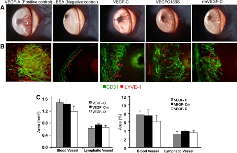
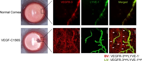
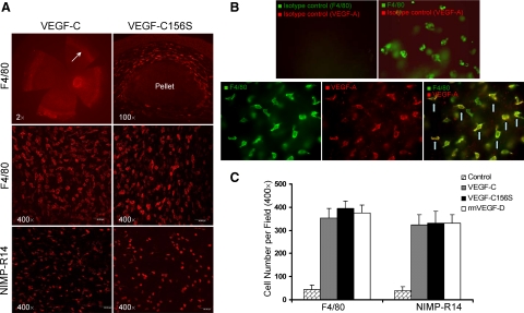
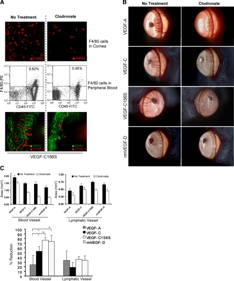
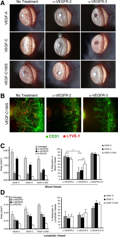
Vascular endothelial growth factor receptor (VEGFR)-2 is a major stimulator of hemangiogenesis (HA), whereas VEGFR-3 stimulates lymphangiogenesis (LA). Contrary to this understanding, we demonstrate that implantation of pellets containing VEGFR-3-specific ligands (VEGF-C156S and recombinant murine VEGF-D) into the corneal stroma induce not only LA but also robust HA characterized by blood vessels that are positive for VEGFR-3 expression. The implantation of pellets containing VEGFR-3-specific ligands also leads to the recruitment of VEGF-A-secreting macrophages. Depletion of these infiltrating macrophages using clodronate-liposome administration shows a significant reduction in HA as well as LA. Blockade of either VEGFR-2 or VEGFR-3 signaling reduces both HA and LA; however, the percent reduction of HA is greater in the VEGFR-2 blockade group. In addition, in the VEGFR-3 blockade group, the percent reduction of HA is significantly greater with VEGFR-3-specific ligands than that by VEGF-A or VEGF-C. Collectively, our data suggest that VEGFR-3-specific signaling can induce new blood vessels, to which macrophages contribute a major role, and signify its potential as an additional therapeutic target to the existing VEGF-A/VEGFR-2 signaling-based antiangiogenesis strategies.


Figures

Figure 1
Figure 1
Corneal blood and lymph vessel growth by VEGFR-3-specific stimulation via VEGF-C156S or rmVEGF-D pellet implants. A: Pellets containing 80 ng of VEGF-C156S or rmVEGF-D induced comparable neovascularization to VEGF-C as detected by biomicroscopy. Pellets containing VEGF-A and BSA were implanted as positive and negative controls, respectively. B: Immunohistochemical staining of flat mount corneas (×100 magnification) revealed similar densities of CD31highLYVE-1 blood vessels (green) and CD31lowLYVE-1high (red) lymph vessels in VEGF-C, VEGF-C156S (VEGF-Cm), and rmVEGF-D pellet-implanted corneas. The growth of the vessels can be seen from the left toward the pellets implanted in the avascular cornea, toward the right of the figures. C: For quantitative comparison of HA and LA induced by different ligands, fluorescent micrographs under low magnification power (×2) were analyzed with NIH software (Image J 1.34). The area of vessels was measured in mm2 and then analyzed as percentage of the total corneal area covered by vessels. Graphs represent mean values (±SEM) of five mice in each group. No significant difference was observed in the area covered by either blood vessels or lymphatic vessels among the different VEGF pellet-implanted groups.
Figure 2
Figure 2
Expression of VEGFR-3 by VEGF-C156S induced new blood vessels. A representative portion (boxed area) of newly formed vessels invading the cornea after 1 week post-VEGF-C156S pellet implantation, and the same area from the normal cornea without pellet implantation was photographed after double immunostaining of the whole-mount cornea with VEGFR-3 (red) and LYVE-1 (green). Merged images of LYVE-1 and VEGFR-3 represent lymphatic vessels (VEGFR-3highLYVE-1high) in yellow-green color, whereas LYVE-1 tubular structures in red color (marked with white arrows) represent VEGFR-3-positive blood vessels. Negative controls stained with isotype-matched antibodies showed no staining.
Figure 3
Figure 3
Abundant numbers of F4/80+ macrophages and NIMP-R14+ neutrophils are recruited to the corneal stroma. A: Fluorescent micrographs of immunohistochemically stained (F4/80 or NIMP-R14) flat-mount corneas showed that abundant numbers of innate immune cells are recruited to the corneal stroma, especially near the limbus (marked with arrows) and around the pellet. Two weeks after implantation of VEGF-C and VEGF-C156S pellets, F4/80+ macrophages reached peak levels of infiltration. Neutrophils identified with the specific marker NIMP-R14 were relatively smaller in size and showed maximal infiltration at week 1 postimplantation of VEGF-C and VEGF-C156S pellets. B: Double staining of F4/80 with VEGF-A after VEGF-C156S pellet implantation showed that a majority of the F4/80+ cells (green) are costained with VEGF-A (red) as shown in merged images (yellow; marked with arrows). C: F4/80+ and NIMP-R14+ cells were counted manually under high magnification power (×400) confocal fluorescein micrographs at 2 and 1 week, respectively. Graphs represent mean values (±SEM) of five mice in each group, and no significant difference in the numbers of infiltrating cells was found among different VEGF pellet-implanted groups.
Figure 4
Figure 4
Antiangiogenic effects of macrophage depletion. A: Intraperitoneal injection of clodronate effectively depleted the F4/80+ cells both locally in cornea (IHC, panel 1) and systemically in peripheral blood (FACS plots, panel 2). Representative micrographs (panel 3) taken 1 week after VEGF-C156S pellet implantation showed significant reduction of both blood vessels (CD31highLYVE-1, green) and lymphatic vessels (CD31lowLYVE-1high, red) in clodronate liposome-treated eyes as compared with untreated eyes. B: Slit lamp photographs taken 1 week after pellet implantation showed that systemic macrophage depletion by CL2MDP-lip has an inhibitory effect on corneal vasculogenesis induced by all four proangiogenic growth factors: VEGF-A, VEGF-C, VEGF-C156S, and rmVEGF-D. C: The area of blood vessels and lymphatic vessels was measured in mm2 and then analyzed as the percent reduction in vessel area by comparing to the mean values of vessel area in control groups (no treatment). The percent reduction of blood vessels was significantly higher in the VEGF-C156S and rmVEGF-D groups as compared with those in the VEGF-A and VEGF-C. The difference in percent reduction of lymphatic vessels was not statistically significant among the different VEGF-implanted groups (*P < 0.05). Graphs represent mean values (±SEM) of five mice in each group.
Figure 5
Figure 5
Antiangiogenic effects of VEGFR blockade. Systemic administration of either anti-VEGFR-2 (α-VEGFR-2)- or anti-VEGFR-3 (α-VEGFR-3)-blocking antibodies inhibit formation of both blood vessels and lymphatic vessels. A: Slit lamp photographs taken 1 week after corneal pellet implantation in control and treatment groups show that neutralization of either VEGFR-2 or VEGFR-3 causes suppression of neovascularization, although the inhibitory effects were more evident in eyes treated with VEGFR-2 blockade. B: Representative immunofluorescence photomicrographs (×100 magnification) taken 1 week after VEGF-C156S pellet implantation with or without treatment confirm that both VEGFR-2 and VEGFR-3 have an inhibitory effect on both blood vessels (green) and lymphatic vessels (red). C: The area of blood vessels was measured in mm2 and then analyzed as the percent reduction in vessel area by comparing to the mean values of vessel area in untreated group. Suppression of HA in VEGFR-2 blockade-treated eyes was significantly greater in VEGF-A and VEGF-C induced neovascularization as confirmed by morphometric analysis. The difference in the inhibition of VEGF-C156S induced HA between VEGFR-2 blockade- and VEGFR-3 blockade-treated eye was not statistically significant. D: The area of lymphatic vessels was measured in mm2 and then analyzed as the percent reduction in vessel area by comparing to the mean values of vessel area in untreated group. Inhibition of LA was not significantly different between the VEGFR-2 and VEGFR-3 blockade-treated groups (*P < 0.007; **P < 0.05). Graphs represent mean values (±SEM) of five mice in each group.

References

    1. Ferrara N, Davis-Smyth T. The biology of vascular endothelial growth factor. Endocr Rev. 1997;18:4–25. - PubMed
    1. Shibuya M, Ito N, Claesson-Welsh L. Structure and function of vascular endothelial growth factor receptor-1 and -2. Curr Top Microbiol Immunol. 1999;237:59–83. - PubMed
    1. Alitalo K, Carmeliset P. Molecular mechanism of lymphangiogenesis in health and disease. Cancer Cell. 2002;1:219–227. - PubMed
    1. Shibuya M, Claesson-Welsh L. Signal transduction by VEGF receptors in regulation of angiogenesis and lymphangiogenesis. Exp Cell Res. 2005;312:549–560. - PubMed
    1. Cao Y, Linden P, Farnebo J, Cao R, Eriksson A, Kumar V, Qi JH, Claesson-Welsh L, Alitalo K. Vascular endothelial growth factor C induces angiogenesis in vivo. Proc Natl Acad Sci USA. 1998;95:14389–14394. - PMC - PubMed

Publication types

MeSH terms

Substances

LinkOut - more resources