. 2009 Dec 4;284(49):33949-56.
doi: 10.1074/jbc.M109.070201. Epub 2009 Oct 5.

A role for the ATP7A copper-transporting ATPase in macrophage bactericidal activity

Affiliations

A role for the ATP7A copper-transporting ATPase in macrophage bactericidal activity

Carine White et al. J Biol Chem. .

Abstract

Copper is an essential micronutrient that is necessary for healthy immune function. This requirement is underscored by an increased susceptibility to bacterial infection in copper-deficient animals; however, a molecular understanding of its importance in immune defense is unknown. In this study, we investigated the effect of proinflammatory agents on copper homeostasis in RAW264.7 macrophages. Interferon-gamma was found to increase expression of the high affinity copper importer, CTR1, and stimulate copper uptake. This was accompanied by copper-stimulated trafficking of the ATP7A copper exporter from the Golgi to vesicles that partially overlapped with phagosomal compartments. Silencing of ATP7A expression attenuated bacterial killing, suggesting a role for ATP7A-dependent copper transport in the bactericidal activity of macrophages. Significantly, a copper-sensitive mutant of Escherichia coli lacking the CopA copper-transporting ATPase was hypersensitive to killing by RAW264.7 macrophages, and this phenotype was dependent on ATP7A expression. Collectively, these data suggest that copper-transporting ATPases, CopA and ATP7A, in both bacteria and macrophage are unique determinants of bacteria survival and identify an unexpected role for copper at the host-pathogen interface.


Figures
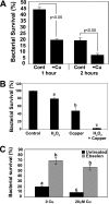
FIGURE 1.
FIGURE 1.
Copper enhances bacterial killing by RAW264.7 macrophage cells. A, RAW264.7 macrophages were treated with 25 ng/ml IFN-γ in the presence or absence of 20 μm copper prior to exposure to E. coli for 30 min to allow phagocytosis to occur. After removal of extracellular bacteria, bacterial survival was measured following incubations for 1 or 2 h at 37 °C and expressed as a percentage of initial internalized E. coli (mean ± S.D.; n = 3; p < 0.05, Student's t test). B, in vitro bacterial survival was assessed following exposure of E. coli to 0.5 mm H2O2, 10 μm CuCl2 or H2O2 plus CuCl2. Bacterial survival rates were calculated and expressed as percentage survival compared with the untreated group (mean ± S.D.; n = 3; p < 0.05). Different letters signify values of significant difference (Tukey's post hoc analysis). C, bacterial survival was assayed after 2 h in IFN-γ-treated RAW264.7 macrophages in the presence or absence of 20 μm copper and a 50 μm concentration of the antioxidant ebselen (mean ± S.D.; n = 3; p < 0.05, one- and two-way analysis of variance). Different letters signify values of significant difference (Tukey's post hoc analysis).
FIGURE 2.
FIGURE 2.
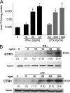
Proinflammatory agents stimulate copper uptake and CTR1 expression in RAW264.7 cells. A, copper uptake activity. RAW264.7 cells were pretreated with serum-free medium with or without IFN-γ or LPS at the indicated concentrations for 24 h, and copper uptake in the presence of 1 μm 64Cu was measured over 5 min and expressed per total protein concentration (mean ± S.D.; n = 3; *, p < 0.05, Student's t test). B, Western blot analysis of CTR1 protein levels in total cell lysates of RAW264.7 macrophage cells exposed for 24 h to a range of IFN-γ and LPS concentrations. Relative band intensities against tubulin are shown in parentheses.
FIGURE 3.
FIGURE 3.
Proinflammatory agents stimulate ATP7A expression in RAW264.7 cells. Western blot analysis of ATP7A protein levels in total cell lysates of RAW264.7 macrophage cells exposed for 24 h to a range of IFN-γ (A) and LPS concentrations (B). Relative band intensities against tubulin are shown in parentheses.
FIGURE 4.
FIGURE 4.
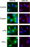
Lipopolysaccharide and IFN-γ induce trafficking of ATP7A in RAW264.7 cells. Shown is immunofluorescence analysis of ATP7A protein in RAW264.7 cells grown for 24 h in the presence of 100 ng/ml LPS, 25 ng/ml IFN-γ, or 20 μm CuCl2. Cells were fixed, permeabilized, and probed with antibodies against ATP7A and anti-rabbit antibodies conjugated to Alexa-488 (green) or antibodies against the Golgi marker GM130 and anti-mouse antibodies conjugated to Alexa-594 (red). Nuclei were labeled with 4′,6-diamidino-2-phenylindole (blue).
FIGURE 5.
FIGURE 5.
The trafficking of ATP7A in response to IFN-γ is copper-dependent. A, IFN-γ-induced trafficking of ATP7A is inhibited by the copper chelator TTM. RAW264.7 cells were cultured for 24 h in the presence of IFN-γ with or without 10 nm TTM. ATP7A protein was detected by immunofluorescence microscopy after cells were fixed, permeabilized, and probed with antibodies against ATP7A and anti-rabbit antibodies conjugated to Alexa-488 (green). B, tetrathiomolybdate does not inhibit the increase in ATP7A protein expression during inflammation. RAW264.7 were treated for 24 h with serum-free medium alone or the same medium containing 100 ng/ml LPS or 25 ng/ml IFN-γ in the presence or absence of 10 nm TTM. ATP7A protein was detected in total cell lysates by Western blot analysis. Relative band intensities against tubulin are shown in parentheses.
FIGURE 6.
FIGURE 6.
Time course analysis of IFN-γ-stimulated ATP7A trafficking and expression. Shown are immunofluorescence analysis (A) and Western blot analysis (B) of ATP7A protein in RAW264.7 cells exposed to 25 ng/ml IFN-γ for the indicated times. Relative band intensities against tubulin are shown in parentheses.
FIGURE 7.
FIGURE 7.
IFN-γ stimulates partial trafficking of the ATP7A protein to the phagosomal compartment in RAW264.7 macrophages. A, partial co-localization of ATP7A with phagosomal membranes. RAW264.7 macrophages were stimulated overnight with IFN-γ and allowed to phagocytose latex beads to allow identification of the phagosomal compartments (Bright field). B, ATP7A was detected by immunofluorescence microscopy in the same cells as in A. C, co-fractionation of ATP7A with the phagosomal membrane marker Lamp-1. IFN-γ-stimulated RAW264.7 cells were allowed to phagocytose latex beads, and the phagosomal compartment was isolated via subcellular fractionation. The bead fraction and total protein fraction were subjected to SDS-PAGE. Immunoblot analysis revealed abundant ATP7A in Lamp-1-positive bead fraction, which lacked the Golgi marker protein GM130. Note that Lamp-1 was poorly detected in total lysates. D, partial co-localization of ATP7A with Rab7-Q67L in IFN-γ-stimulated RAW264.7 cells. Cells were transiently transfected with green fluorescent protein-tagged Rab7-Q67L plasmid and then exposed for 24 h to basal medium or medium containing 25 ng/ml IFN-γ. Cells were fixed, permeabilized, and probed with antibodies against ATP7A followed by anti-rabbit antibodies conjugated to Alexa-594.
FIGURE 8.
FIGURE 8.
E. coli survival is dependent on ATP7A expression in RAW264.7 macrophages. A, immunoblot analysis of ATP7A protein in RAW264.7 cells stably transfected with either ATP7A RNAi or control RNAi. B, Western blot analysis of ceruloplasmin protein in concentrated conditioned medium collected from ATP7A RNAi and control RNAi cells treated for 24 h in the presence or absence of 25 ng/ml IFN-γ. Note that ceruloplasmin basal expression or induction by IFN-γ is not impaired by ATP7A depletion. C, ceruloplasmin activity was assayed from concentrated conditioned medium from control RNAi or ATP7A RNAi macrophages treated for 24 h in the presence or absence of 20 μm CuCl2. Values were normalized against total protein content of each sample (mean ± S.D.; n = 3; two-way analysis of variance). Note the suppression of ceruloplasmin activity in ATP7A-depleted cells and the rescue by the addition of copper. D, survival of wild type E. coli in control RNAi- or ATP7A RNAi-transfected RAW264.7 cells pretreated overnight with 25 ng/ml IFN-γ in the presence or absence of 20 μm CuCl2. Note that bacterial survival was significantly increased in ATP7A-depleted cells compared with control cells and that copper pretreatment of ATP7A-depleted cells restored bactericidal activity to control levels (mean ± S.D.; n = 3; two-way analysis of variance).
FIGURE 9.
FIGURE 9.
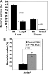
The bacterial copper exporter, CopA, is a determinant of E. coli survival in RAW264.7 macrophages. A, the survival of wild type (Wt) and ΔcopA deletion mutant E. coli in IFN-γ stimulated RAW264.7 macrophages. The percentage survival data is shown for 1 and 2 h kill times (postuptake) (mean ± S.D.; n = 3). B, the killing of ΔcopA mutant E. coli is attenuated in ATP7A-depleted RAW264.7 macrophages. ATP7A RNAi- and control RNAi-transfected cells were stimulated overnight with IFN-γ (mean ± S.D.; n = 3).

References

    1. Kim B. E., Nevitt T., Thiele D. J. (2008) Nat. Chem. Biol. 4, 176–185 - PubMed
    1. Kadiiska M. B., Mason R. P. (2002) Spectrochim. Acta A Mol. Biomol. Spectrosc. 58, 1227–1239 - PubMed
    1. Jiménez Del Río M., Vélez-Pardo C. (2004) Arch. Med. Res. 35, 185–193 - PubMed
    1. Wardman P., Candeias L. P. (1996) Radiat. Res. 145, 523–531 - PubMed
    1. Prohaska J. R., Lukasewycz O. A. (1981) Science 213, 559–561 - PubMed

Publication types