. 2010 Jan;59(1):128-34.
doi: 10.2337/db09-1098. Epub 2009 Oct 6.

ATP-sensitive K+ channel mediates the zinc switch-off signal for glucagon response during glucose deprivation

Affiliations

ATP-sensitive K+ channel mediates the zinc switch-off signal for glucagon response during glucose deprivation

Michela Slucca et al. Diabetes. 2010 Jan.

Abstract

Objective: The intraislet insulin hypothesis proposes that glucagon secretion during hypoglycemia is triggered by a decrease in intraislet insulin secretion. A more recent hypothesis based on in vivo data from hypoglycemic rats is that it is the decrease in zinc cosecreted with insulin from beta-cells, rather than the decrease in insulin itself, that signals glucagon secretion from alpha-cells during hypoglycemia. These studies were designed to determine whether closure of the alpha-cell ATP-sensitive K(+) channel (K(ATP) channel) is the mechanism through which the zinc switch-off signal triggers glucagon secretion during glucose deprivation.

Research design and methods: All studies were performed using perifused isolated islets.

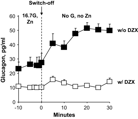
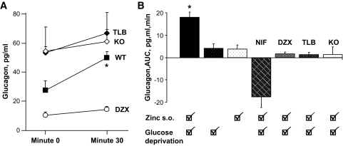
Results: In control experiments, the expected glucagon response to an endogenous insulin switch-off signal during glucose deprivation was observed in wild-type mouse islets. In experiments with streptozotocin-treated wild-type islets, a glucagon response to an exogenous zinc switch-off signal was observed during glucose deprivation. However, this glucagon response to the zinc switch-off signal during glucose deprivation was not seen in the presence of nifedipine, diazoxide, or tolbutamide or if K(ATP) channel knockout mouse islets were used. All islets had intact glucagon responses to epinephrine.

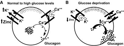
Conclusions: These data demonstrate that closure of K(ATP) channels and consequent opening of calcium channels is the mechanism through which the zinc switch-off signal triggers glucagon secretion during glucose deprivation.


Figures

FIG. 1.
FIG. 1.
A. Endogenous insulin switch-off signal to α-cells in WT islets. Isolated islets were exposed to 16.7 mmol/l glucose (16.7G) for 30 min (from −30 to 0 min). At min 0 the perifusion was either changed to 0 glucose or not changed for the ensuing 30 min. The closed circles at times −10 to 0 min represent the average of experimental and control values when islets were exposed to 16.7 mmol/l glucose. Insulin secretion declined when glucose infusion was changed to 0 mmol/l at time 0 min, generating an endogenous insulin switch-off signal from β-cells to neighboring α-cells (closed boxes). Insulin levels did not decrease significantly when the glucose perifusion was not changed (closed diamonds). Results are expressed as mean ± SE of eight replicate perifusions in experimental studies (closed boxes) and seven replicate perifusions in control studies (closed diamonds). See text for statistical information. A and B: Glucagon responses to an endogenous insulin switch-off signal during perifusion of wild-type mouse islets. Glucagon levels increased significantly only when endogenous insulin secretion (A) was switched-off in response to glucose deprivation at time 0 min (■, n = 8). No glucagon response was observed if the endogenous insulin switch-off signal was absent (♦, n = 7). ● at times −10 to 0 min represent the average of experimental and control values. See text for statistical information.
FIG. 2.
FIG. 2.
Glucagon response in the presence and absence of an exogenous zinc switch-off signal during perifusion of STZ-induced diabetic wild-type mouse islets. Islets were pretreated with STZ to kill β-cells, thereby preventing endogenous insulin and zinc secretion. After an initial 30-min perifusion with 16.7 mmol/l glucose and zinc (Zn), at time 0 min the perifusate was changed. Glucagon levels increased significantly only when a perifusate containing no glucose and no zinc, which generated an exogenous zinc switch-off signal to α-cells during glucose deprivation, was begun at 0 min (■, n = 5). Glucagon secretion did not increase if at time 0 min only glucose, but not zinc, was switched off (○, n = 4) or if only zinc but not glucose was switched off (●, n = 4). If at time 0 min nifedipine (NIF) was added to the perifusate, glucagon secretion not only failed to rise but was suppressed despite the presence of the zinc switch-off signal during glucose deprivation (□, n = 3). ▴ at times −10 to 0 min represent the average of experimental values and control values.
FIG. 3.
FIG. 3.
Glucagon responses from STZ-induced diabetic wild-type mouse islets to an exogenous zinc switch-off signal during glucose deprivation in the presence of diazoxide. After an initial 30-min perifusion with 16.7 mmol/l glucose, diazoxide (DZX), and zinc, both glucose and zinc were discontinued at time 0 min and only diazoxide was continued for the last 30 min. Diazoxide prevented any significant increase in glucagon secretion despite the presence of a zinc switch-off signal during glucose deprivation (□, n = 3). For the purpose of comparison, ■ are the same data reported in Fig. 2 and represent the glucagon response to a zinc switch-off signal during glucose deprivation in the absence of diazoxide infusion. See text for statistical information.
FIG. 4.
FIG. 4.
Glucagon responses from STZ-induced diabetic wild-type mouse islets to an exogenous zinc switch-off signal during glucose deprivation in the presence of tolbutamide (50 μmol/l). After an initial 30-min perifusion with 16.7 mmol/l glucose, tolbutamide (TLB), and zinc, both glucose and zinc were discontinued at time 0 min and only tolbutamide was continued for the last 30 min. Tolbutamide prevented any significant increase in glucagon secretion despite the presence of a zinc switch-off signal during glucose deprivation (□, n = 3). For the purpose of comparison, ■ are the same data reported in Fig. 2 and represent the glucagon response to a zinc switch-off signal during glucose deprivation in the absence of tolbutamide infusion. The glucagon response is expressed as percentage of baseline because of the higher glucagon baseline in the islets perifused with tolbutamide due to tolbutamide-mediated closure of the KATP channels. See text for statistical information.
FIG. 5.
FIG. 5.
Glucagon responses in the presence of an exogenous zinc switch-off signal during perifusion of STZ-induced diabetic SUR1KO mouse islets. STZ-induced diabetic SUR1KO mouse islets were perifused with 16.7 mmol/l glucose and zinc for 30 min. Then, at time 0 min, both glucose and zinc were discontinued. Glucagon secretion from SUR1KO mouse islets failed to increase despite the presence of a zinc switch-off signal during glucose deprivation (□, n = 4). For the purpose of comparison, ■ are the same data reported in Fig. 2 and represent the glucagon rise in response to a zinc switch-off signal during glucose deprivation in STZ-induced diabetic wild-type mouse islets. The glucagon response is expressed as a percentage of baseline because of the higher glucagon baseline in SUR1KO mouse due to the absence of KATP channels. See text for statistical information.
FIG. 6.
FIG. 6.
Summary of glucagon responses. A: Comparison between glucagon values baseline at time 0 min immediately before the zinc switch-off signal during glucose deprivation and at time +30 min (means ± SE). Compared with the untreated wild-type islets, baseline glucagon levels at time 0 min were higher in the depolarized state (TLB, KO) and lower in the polarized state (DZX), all P < 0.05. At time +30 min, no differences were noted among the groups. A significant difference was noted when comparing time 0 to time +30 min in the untreated wild-type islet group (P < 0.02). B: Glucagon responses expressed as area under the curve (AUC) after zinc switch-off signal. A statistically significant (P < 0.001) glucagon response occurred only when a zinc switch-off signal was provided during glucose deprivation in wild-type islets pretreated with STZ.
FIG. 7.
FIG. 7.
Glucagon responses to epinephrine. Glucagon responses to epinephrine were intact in both wild-type and SUR1KO islets. ♦, ♢, and hatched diamonds represent data obtained, respectively, from wild-type islets, STZ-induced diabetic wild-type islets, and STZ-induced diabetic SUR1KO islets. The glucagon response correlated significantly with the prestimulated glucagon level.
FIG. 8.
FIG. 8.
Regulation of glucagon secretion by zinc. A: α-Cell electrical and hormonal status in states of physiologic and elevated glucose conditions. β-Cells release zinc and insulin hexamers into the intraislet periportal circulation. Zinc dissociates from insulin and reaches downstream α-cells, where it binds to and opens the KATP channels. K+ ions leave the cell and hyperpolarize the α-cell, thus preventing voltage-dependent calcium channels from opening. Glucagon granules are not mobilized and remain stored inside the cell. B: When blood glucose levels decrease in response to hypoglycemic levels, β-cell insulin and zinc secretion decrease as well. KATP channels on α-cells close, K+ remains in the cell, and the α-cell depolarizes, which induces calcium channels to open and calcium enters the cell. Intracellular calcium rises, inducing glucagon exocytotic granules to migrate to the plasma membrane, where they fuse and release glucagon into the portal venous system.

References

    1. Banarer S, McGregor VP, Cryer PE: Intraislet hyperinsulinemia prevents the glucagon response to hypoglycemia despite an intact autonomic response. Diabetes 2002;51:958–965 - PubMed
    1. Zhou H, Tran PO, Yang S, Zhang T, LeRoy E, Oseid E, Robertson RP: Regulation of alpha-cell function by the beta-cell during hypoglycemia in wistar rats: the “switch-off” hypothesis. Diabetes 2004;53:1482–1487 - PubMed
    1. Hope KM, Tran PO, Zhou H, Oseid E, Leroy E, Robertson RP: Regulation of α-cell function by the β-cell in isolated human and rat islets deprived of glucose: the “switch-off” hypothesis. Diabetes 2004;53:1488–1495 - PubMed
    1. Samols E, Stagner JI, Ewart RB, Marks V: The order of islet microvascular cellular perfusion is β-α-δ in the perfused rat pancreas. J Clin Invest 1988;82:350–353 - PMC - PubMed
    1. Rorsman P, Berggren PO, Bokvist K, Ericson H, Mohler H, Ostenson CG, Smith PA: Glucose-inhibition of glucagon secretion involves activation of gabaa-receptor chloride channels. Nature 1989;341:233–236 - PubMed

Publication types

MeSH terms